Ann Clin Microbiol 2024;27:149-153. Can Acanthamoeba keratitis be properly diagnosed without culture in the real-world clinical microbiology laboratory?: a case report
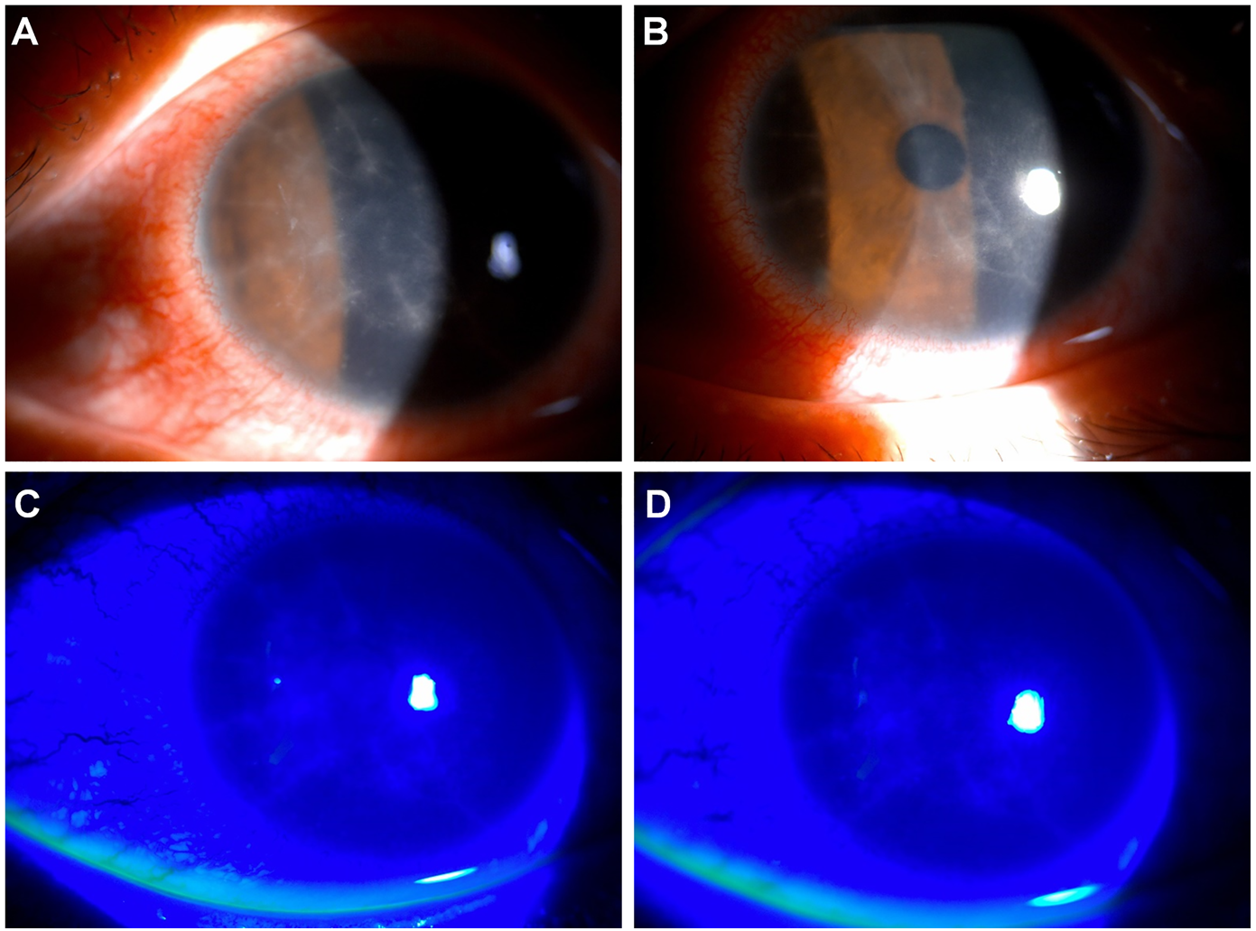
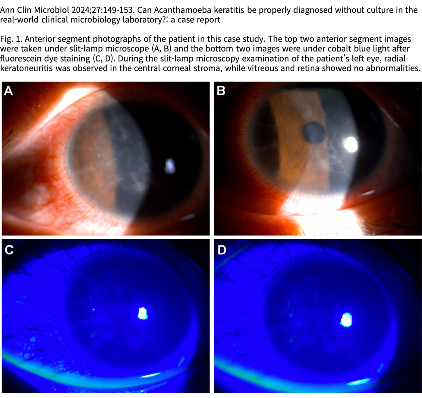
Fig. 1. Anterior segment photographs of the patient in this case study. The top two anterior segment images were taken under slit-lamp microscope (A, B) and the bottom two images were under cobalt blue light after fluorescein dye staining (C, D). During the slit-lamp microscopy examination of the patient’s left eye, radial keratoneuritis was observed in the central corneal stroma, while vitreous and retina showed no abnormalities.