Ann Clin Microbiol 2024;27:149-153. Can Acanthamoeba keratitis be properly diagnosed without culture in the real-world clinical microbiology laboratory?: a case report
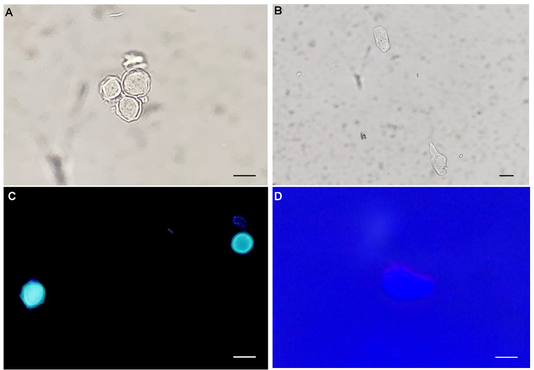
Fig. 2. Morphological analysis of Acanthamoeba spp. using calcofluor-white staining. (A) Acanthamoeba cysts in a group under light microscopy (400×, scale bar = 10 μm). (B) Acanthamoeba trophozoites with a single large nucleus and numerous vacuoles were conspicuous under light microscopy (200×, scale bar = 10 μm). (C) Remarkable Acanthamoeba cysts with double cyst walls under fluorescence microscopy (400×, scale bar = 10 μm). (D) Acanthamoeba trophozoites were relatively unremarkable under fluorescence microscopy (400×, scale bar = 10 μm).
