Myeong Hee Kim![]()

1. Gushiken AC, Saharia KK, Baddley JW. Cryptococcosis. Infect Dis Clin North Am 2021;35:493-514.

2. Maziarz EK, Perfect JR. Cryptococcosis. Infect Dis Clin North Am 2016;30:179-206. 
3. Speed B, Dunt D. Clinical and host differences between infections with the two varieties of Cryptococcus neoformans. Clin Infect Dis 1995;21:28-34.
4. Galanis E, MacDougall L, Kidd S, Morshed M; British Columbia Cryptococcus gattii Working Group. Epidemiology of Cryptococcus gattii, British, Columbia, Canada, 1999-2007. Emerg Infect Dis 2010;16:251-7.
5. Mitchell DH, Sorrell TC, Allworth AM, Heath CH, McGregor AR, Papanaoum K, et al. Cryptococcal disease of the CNS in immunocompetent hosts: influence of cryptococcal variety on clinical manifestations and outcome. Clin Infect Dis 1995;20:611-6.
6. May RC, Stone NRH, Wiesner DL, Bicanic T, Nielsen K. Cryptococcus: from environmental saprophyte to global pathogen. Nat Rev Microbiol 2016;14:106-17.
7. Datta K, Bartlett KH, Marr KA. Cryptococcus gattii: emergence in western North America: exploitation of a novel ecological niche. Interdiscip Perspect Infect Dis 2009;2009:176532.
8. Kidd SE, Hagen F, Tscharke RL, Huynh M, Bartlett KH, Fyfe M, et al. A rare genotype of Cryptococcus gattii caused the cryptococcosis outbreak on Vancouver Island (British Columbia, Canada). Proc Natl Acad Sci USA 2004;101:17258-63.
9. Chen J, Varma A, Diaz MR, Litvintseva AP, Wollenberg KK, Kwon-Chung KJ. Cryptococcus neoformans strains and infection in apparently immunocompetent patients, China. Emerg Infect Dis 2008;14:755-62.
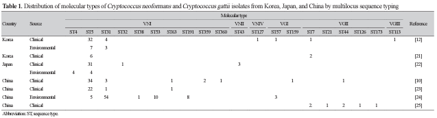
10. Wu SY, Lei Y, Kang M, Xiao YL, Chen ZX. Molecular characterisation of clinical Cryptococcus neoformans and Cryptococcus gattii isolates from Sichuan province, China. Mycoses 2015;58:280-7.
11. Kaocharoen S, Ngamskulrungroj P, Firacative C, Trilles L, Piyabongkarn D, Banlunara W, et al. Molecular epidemiology reveals genetic diversity amongst isolates of the Cryptococcus neoformans/C. gattii species complex in Thailand. PLoS Negl Trop Dis 2013;7:e2297.
12. Park SH, Choi SC, Lee KW, Kim MN, Hwang SM. Genotypes of clinical and environmental isolates of Cryptococcus neoformans and Cryptococcus gattii in Korea. Mycobiology 2015;43:360-5.
13. Hwang SM. Molecular typing of clinical Cryptococcus gattii isolates in Korea. J Bacteriol Virol 2012;42:152-5.
14. Chang CC, Harrison TS, Bicanic TA, Chayakulkeeree M, Sorrell TC, Warris A, et al. Global guideline for the diagnosis and management of cryptococcosis: an initiative of the ECMM and ISHAM in cooperation with the ASM. Lancet Infect Dis 2024;24:e495-512.
15. Park SW, Choi JY, Kim AY, Chang DS. A case of cryptococcal abscess involving deep neck space in an immunocompetent patient. Korean J Otorhinolaryngol-Head Neck Surg 2011;54:638-41.
16. Choo MJ, Shin SO, Yang SK, Jin HR. Cryptococcal infection combined with cholesteatoma. Korean J Otolaryngol-Head Neck Surg 1999;42:639-42.
17. Hyun JJ, Choi JH, Park S, Jeong HW, Jung SJ, Kee SY, et al. A case report on cryptococcal lymphadenitis in an immunocompetent adult patient. Infect Chemother 2005;37:350-4.
18. Takashima M and Sugita T. Taxonomy of pathogenic yeasts Candida, Cryptococcus, Malassezia, and Trichosporon. Med Mycol J 2022;63:119-32.
19. Meyer W, Aanensen DM, Boekhout T, Cogliati M, Diaz MR, Esposto MC, et al. Consensus multi-locus sequence typing scheme for Cryptococcus neoformans and Cryptococcus gattii. Med Mycol 2009;47:561-70.
20. Khayhan K, Hagen F, Pan W, Simwami S, Fisher MC, Wahyuningsih R, et al. Geographically structured populations of Cryptococcus neoformans variety grubii in Asia correlate with HIV status and show a clonal population structure. PLoS One 2013;8:e72222.
21. Choi YH, Ngamskulrungroj P, Varma A, Sionov E, Hwang SM, Carriconde F, et al. Prevalence of the VNIc genotype of Cryptococcus neoformans in non-HIV-associated cryptococcosis in the Republic of Korea. FEMS Yeast Res 2010;10:769-78.
22. Mihara T, Izumikawa K, Kakeya H, Ngamskulrungroj P, Umeyama T, Takazono T, et al. Multilocus sequence typing of Cryptococcus neoformans in non-HIV associated cryptococcosis in Nagasaki, Japan. Med Mycol 2013;51:252-60.
23. Dou HT, Xu YC, Wang HZ, Li TS. Molecular epidemiology of Cryptococcus neoformans and Cryptococcus gattii in China between 2007 and 2013 using multilocus sequence typing and the DiversiLab system. Eur J Clin Microbiol Infect Dis 2015;34:753-62.
24. Dou H, Wang H, Xie S, Chen X, Xu Z, Xu Y. Molecular characterization of Cryptococcus neoformans isolated from the environment in Beijing, China. Med Mycol 2017;55:737-47.
25. Zang X, Ke W, Wang L, Wu H, Huang Y, Deng H, et al. Molecular epidemiology and microbiological characteristics of Cryptococcus gattii VGII isolates from China. PLoS Negl Trop Dis 2022;16:e0010078.
26. Zaragoza O, Rodrigues ML, De Jesus M, Frases S, Dadachova E, Casadevall A. The capsule of the fungal pathogen Cryptococcus neoformans. Adv Appl Microbiol 2009;68:133-216.
27. Shin JH. Laboratory diagnosis of opportunistic fungal infections. Ann Clin Microbiol 1998;1:37-43.
28. de Repentigny L. Serodiagnosis of candidiasis, aspergillosis, and cryptococcosis. Clin Infect Dis 1992;14(suppl 1):S11-22.
29. Williams DA, Kiiza T, Kwizera R, Kiggundu R, Velamakanni S, Meya DB, et al. Evaluation of fingerstick cryptococcal antigen lateral flow assay in HIV-infected persons: a diagnostic accuracy study. Clin Infect Dis 2015;61:464-7.
30. Boulware DR, Rolfes MA, Rajasingham R, von Hohenberg M, Qin Z, Taseera K, et al. Multisite validation of cryptococcal antigen lateral flow assay and quantification by laser thermal contrast. Emerg Infect Dis 2014;20:45-53.
31. Vidal JE and Boulware DR. Lateral flow assay for cryptococcal antigen: an important advance to improve the continuum of HIV care and reduce cryptococcal meningitis-related mortality. Rev Inst Med Trop Sao Paulo 2015;57(suppl 19):38-45.
32. Forrest GN, Bhalla P, DeBess EE, Winthrop KL, Lockhart SR, Mohammadi J, et al. Cryptococcus gattii infection in solid organ transplant recipients: description of Oregon outbreak cases. Transpl Infect Dis 2015;17:467-76.
33. Walsh TJ and Chanock SJ. Diagnosis of invasive fungal infections: advances in nonculture systems. Curr Clin Top Infect Dis 1998;18:101-53.
34. Yeo SF and Wong B. Current status of nonculture methods for diagnosis of invasive fungal infections. Clin Microbiol Rev 2002;15:465-84.
35. Tansarli GS and Chapin KC. Diagnostic test accuracy of the BioFire FilmArray meningitis/encephalitis panel: a systematic review and meta-analysis. Clin Microbiol Infect 2020;26:281-90.
36. Ramachandran PS, Cresswell FV, Meya DB, Langelier C, Crawford ED, DeRisi JL, et al. Detection of Cryptococcus DNA by metagenomic next-generation sequencing in symptomatic cryptococcal antigenemia. Clin Infect Dis 2019;68:1978-9.
37. Rogers TR, Verweij PE, Castanheira M, Dannaoui E, White PL, Arendrup MC. Molecular mechanisms of acquired antifungal drug resistance in principal fungal pathogens and EUCAST guidance for their laboratory detection and clinical implications. J Antimicrob Chemother 2022;77:2053-73.
38. Kim SJ, Kwon-Chung KJ, Milne GW, Hill WB, Patterson G. Relationship between polyene resistance and sterol compositions in Cryptococcus neoformans. Antimicrob Agents Chemother 1975;7:99-106.
39. Sanguinetti M, Posteraro B, Sorda ML, Torelli R, Fiori B, Santangelo R, et al. Role of AFR1, an ABC transporter-encoding gene, in the in vivo response to fluconazole and virulence of Cryptococcus neoformans. Infect Immun 2006;74:1352-9.
40. Gerstein AC, Fu MS, Mukaremera L, Li Z, Ormerod KL, Fraser JA, et al. Polyploid titan cells produce haploid and aneuploid progeny to promote stress adaptation. mBio 2015;6:e01340-15.
41. Garcia-Effron G. Rezafungin-mechanisms of action, susceptibility and resistance: similarities and differences with the other echinocandins. J Fungi 2020;6:262.
42. CLSI. Epidemiological cutoff values for antifungal susceptibility testing, M57S. 4th ed. Wayne, PA: Clinical and Laboratory Standards Institute; 2022.
43. European Committee on Antimicrobial Susceptibility Testing. Overview of antifungal ECOFFs and clinical breakpoints for yeasts, moulds and dermatophytes using the EUCAST E.Def 7.4, E.Def 9.4 and E.Def 11.0 procedures. Version 4.0, 2023. https://www.eucast.org/fileadmin/src/media/PDFs/EUCAST_files/AFST/Clinical_breakpoints/EUCAST_BP_ECOFF_v_4.0.pdf [Online] (last visited on 4 October 2024).
44. CLSI. Reference method for broth dilution antifungal susceptibility testing of yeasts, M27-A3. 4th ed. Wayne, PA: Clinical and Laboratory Standards Institute; 2017.
45. Zhang M, Zhou Z, Wang D, Zhou A, Song G, Chen X, et al. Comparative evaluation of Sensititre YeastOne and VITEK 2 against the Clinical and Laboratory Standards Institute M27-E4 reference broth microdilution method for the antifungal susceptibility testing of Cryptococcus neoformans and Cryptococcus gattii. Med Mycol 2022;60:myac009.