Hui-Jin Yu, Tae Yeul Kim, Eun Jeong Won, Hee Jae Huh
Ann Clin Microbiol 2026 June, 29(2):6. Published on 8 May 2026.
Background: To overcome the labor-intensive and technician-dependent nature of the formalin–ether concentration technique (FECT), artificial intelligence (AI)-based automated feces analyzers have been developed for detecting fecal parasites. This study compared the clinical performance of the KU-F40 AI-based automated system with that of FECT in a routine diagnostic environment.
Methods: In total, 1,011 fecal samples were prospectively collected and analyzed concurrently via both FECT and the KU-F40 system. The diagnostic performance of the KU-F40 system was evaluated using FECT as the reference method. The clinical performance parameters included accuracy, Cohen’s kappa coefficient, sensitivity, specificity, positive predictive value (PPV), and negative predictive value (NPV).
Results: The KU-F40 system exhibited an accuracy of 98.5% and Cohen’s kappa of 0.608 relative to FECT. The sensitivity and specificity of the KU-F40 system were 57.1% (95% confidence interval [CI], 34.0–78.2) and 99.4% (95% CI, 98.7–99.8), respectively. When adjusted for an institutional prevalence of 0.45%, its PPV and NPV were 29.9% (95% CI, 15.0–50.7) and 99.8% (95% CI, 99.7–99.9), respectively. Among the 27 parasite-positive samples identified using at least one method, the methods achieved concordant species identification for six samples (22.2%). The KU-F40 system offers superior workflow efficiency by reducing labor time and hazard exposure.
Conclusion: The KU-F40 system demonstrated high specificity and negative predictive value in a low-prevalence setting; however, its limited sensitivity indicates it should not replace conventional examination when clinical suspicion is high. Despite these limitations, it may serve as a useful adjunctive tool that improves laboratory workflow efficiency and minimizes exposure to hazardous reagents.
Yu Jin Park, Xinyi Lu, Sunghyuk Kim, Shinyoung Yoon, Daewon Kim, Ki Ho Hong, Hyukmin Lee, Dongeun Yong
Ann Clin Microbiol 2026 June, 29(2):7. Published on 20 June 2026.
Background: The optimal frequency for repeat antimicrobial susceptibility testing (AST) of isolates of the same species remain undefined despite its importance in antimicrobial therapy. This study assessed suitable repeat AST intervals by analyzing time-dependent changes in susceptibility patterns.
Methods: This retrospective observational study analyzed laboratory AST data collected over 10 years (2010–2019) from a tertiary hospital in Seoul, Korea. We examined AST results for “paired” (same patient, same day) and “successive” (same patient, different times: 1–7, 8–30, and 31–365 days) isolates of common bacterial species: Staphylococcus aureus, Enterococcus faecalis, Enterococcus faecium, Escherichia coli, Klebsiella pneumoniae, Acinetobacter baumannii complex, and Pseudomonas aeruginosa. The essential agreement (EA), categorical agreement (CA), essential MIC increase (EMI), and change from nonresistant to resistant (CNRR and CNRR+EMI) rates were calculated.
Results: Paired isolates showed > 90% CA for most species, although P. aeruginosa had a lower EA (71.5%) and CA (69.6%). For successive isolates, the EMI, CNRR, and CNRR+EMI generally increased over time. The S. aureus and E. faecalis species demonstrated high stability with low CNRR+EMI rates. Conversely, P. aeruginosa exhibited the most notable changes: its CNRR+EMI increased from 4.1% (1–7 days) to 6.6% (31–365 days), and its EMI remained the highest across all time periods, starting at 7.1% within 1–7 days.
Conclusion: The stability of antimicrobial susceptibility patterns varies considerably by bacterial species and time interval. For bacteria with low CNRR+EMI rates, such as S. aureus and E. faecalis, the frequency of repeat AST should be reduced, whereas for bacteria with variable susceptibility patterns, such as P. aeruginosa, more frequent retesting should be performed. The study findings could help laboratories optimize repeat AST protocols and balance timely resistance detection with optimal resource management.
Jeong Su Park, Jehada-Inn Alihuddin, Kidong Kim, Kyoungman Cho, Jung-A Shin, Mulbora Jee
Ann Clin Microbiol 2026 June, 29(2):9. Published on 20 June 2026.
Background: Accurate diagnosis of vulvovaginal candidiasis (VVC) is challenging because of the limitations of conventional wet-mount microscopy, which requires a trained examiner and offers suboptimal sensitivity. This preliminary study evaluated the analytical performance of laser scattering signal amplification (LSSA) combined with deep learning and its preliminary clinical feasibility for detecting Candida culture positivity in women with vaginal symptoms. Methods: LSSA signal data were acquired using the Bacometer from reference strains of five Candida species (10²–105 colony-forming units (CFU)/mL in Luria–Bertani broth). A twostage convolutional neural network (CNN) classifier was developed: Model 1 discriminated negative, C. albicans-positive, and non-albicans-positive specimens; Model 2 distinguished C. albicans from C. tropicalis among the positive specimens. The models were trained on 320 and 80 vials using 60:15:25 stratified splits and 5-fold cross-validation. Twenty vaginal discharge specimens from women with suspected VVC were assessed using fungal culture as the reference standard. Results: Model 1 achieved a hold-out accuracy of 73.8% (95% confidence interval [CI] 62.7–83.0%) for three-class concentration discrimination, whereas Model 2 achieved 77.8% (95% CI 60.8–89.9%) in cascade evaluation. In the pilot clinical evaluation (5 culture-positive and 15 culture-negative specimens), the classifier correctly identified 1 of 5 culture-positive cases (sensitivity 20.0%, 95% CI 0.5–71.6%) and all 15 culture-negative specimens (specificity 100.0%, 95% CI 78.2–100.0%), yielding an overall accuracy of 80.0% (95% CI 56.3–94.3%), positive predictive value (PPV) 100.0% (95% CI 2.5–100.0%), and negative predictive value (NPV) 78.9% (95% CI 54.4–93.9%). Conclusion: This preliminary study observed that concentration- and species-related scattering signals were partially classifiable under controlled reference strain conditions; however, the clinical sensitivity was insufficient (20.0%) in its current form. Correction of the sample pre-processing protocol is required before further clinical evaluation.
Changseung Liu, Young Hee Seo, Kyoung Ho Roh, Hyukmin Lee, Kyungwon Lee
Ann Clin Microbiol 2026 June, 29(2):8. Published on 20 June 2026.
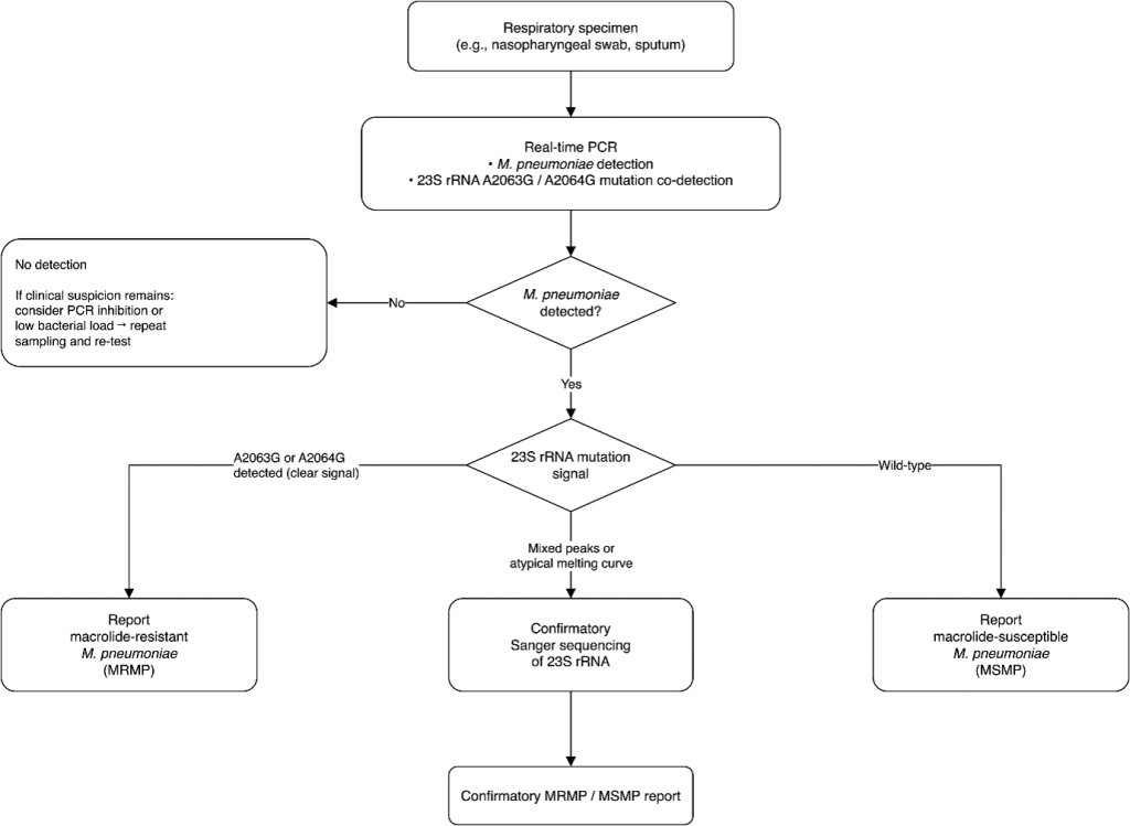
Azithromycin-resistant Neisseria gonorrhoeae poses a threat to the efficacy of gonorrhea treatment. We report the whole-genome characterization of two azithromycin-resistant N. gonorrhoeae ST1600 isolates collected in Busan, South Korea, in 2018 and 2019. Both isolates showed moderate azithromycin resistance (minimum inhibitory concentration 32 µg/mL) mediated by the 23S rRNA C2611T mutation in all four alleles and the -35A deletion in the mtrR promoter, while harboring distinct non-mosaic penA alleles without reduced susceptibility to extended-spectrum cephalosporins. Multilocus sequence typing classified both isolates as ST1600, which is related to ST7363. N. gonorrhoeae multi-antigen sequence typing assigned the 2018 isolate to ST16190, whereas the 2019 isolate represented a novel NG-MAST type closely related to ST16190 (differing by a single SNP in porB). Taken together, these data suggest a limited local transmission cluster with short-term persistence and microevolution, rather than widespread sustained clonal dissemination. Continued phenotypic and genomic surveillance is needed to monitor azithromycin resistance in N. gonorrhoeae and inform national treatment strategies.
Kibum Jeon, Nuri Lee, Hyun Soo Kim, Han-Sung Kim, Wonkeun Song, Jae-Seok Kim
Ann Clin Microbiol 2026 March, 29(1):1. Published on 6 March 2026.
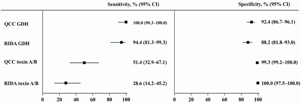
Background: Enzyme immunoassays (EIAs), which detect glutamate dehydrogenase (GDH) and toxin A/B, are widely used to screen for Clostridioides difficile infection (CDI); however, their sensitivity is lower than that of molecular assays. This study aimed to evaluate the performance of two EIAs, C. Diff Quik Chek Complete (QCC) and RIDASCREEN (RIDA), and investigate the cycle threshold (Ct) values from two real-time polymerase chain reaction (PCR) assays (Allplex GI–Bacteria(I) and Xpert C. difficile) in EIA-discordant samples.
Methods: A total of 180 clinical stool samples were tested using QCC, RIDA, and Allplex GI-Bacteria(I) PCR assays. The Xpert C. difficile assay was used to analyze discordant results.
Results: QCC and RIDA showed high sensitivities for GDH detection, 100.0% and 94.4%, respectively. QCC was significantly more sensitive than RIDA for toxin detection (51.4% vs. 28.6%, p = 0.007). In 25 EIA-discordant, Xpert positive samples, the Ct values of the toxin B gene ranged from 31.5 to 44.8 (mean, 38.1) for Allplex PCR and from 23.7 to 36.3 (mean, 30.4) for Xpert PCR. The Ct values of the two PCR assays were not significantly correlated (r = 0.201, p = 0.324).
Conclusion: QCC is a suitable initial immunological test for diagnosing CDI. The lack of correlation in the Ct values between the two real-time PCR assays suggests that assay-specific validation is necessary for cutoff level interpretation.
For Authors
Publisher
Subscribers
for quarterly newsletter subscription & instant discount on publication fee!
Sponsors