
Ann Clin Microbiol 2026;29(3):11. https://doi.org/10.5145/ACM.2026.29.3.11
Received on 08 June 2026, Revised on 21 July 2026, Accepted on 23 July 2026, Published on 22 September 2026.
Copyright © Korean Society of Clinical Microbiology.
This is an Open Access article which is freely available under the Creative Commons Attribution-NonCommercial-NoDerivatives 4.0 International License (CC BY-NC-ND) (https://creativecommons.org/licenses/by-nc-nd/4.0/).
Group A Streptococcus (GAS), or Streptococcus pyogenes, remains predictably susceptible to penicillin, whereas resistance to macrolides has become an important therapeutic and epidemiological concern. This narrative review summarizes the molecular mechanisms, epidemiology, clonal dissemination, and clinical implications of macrolide resistance in GAS. Macrolide resistance is mediated mainly by active efflux, commonly associated with mef genes and the M phenotype, and by ribosomal target-site methylation mediated by erm genes, which produces constitutive or inducible macrolide-lincosamide-streptogramin B resistance. Reported resistance rates vary markedly across geographic regions and study settings, from < 5% in some surveillance populations to > 90% in selected East Asian cohorts. However, direct comparisons are limited by differences in study period, patient population, infection type, and susceptibility testing methods. Molecular epidemiological and whole-genome sequencing studies indicate that both clonal expansion of successful emm lineages and horizontal transfer of resistance determinants carried by mobile genetic elements contribute to dissemination. Clinically, macrolide resistance may compromise treatment in patients requiring non-β-lactam therapy, while erm-mediated cross-resistance can limit the activity of clindamycin in invasive infections. Particular attention is required for inducible clindamycin resistance, which may be missed by routine susceptibility testing. GAS has not developed widespread clinically established penicillin resistance, although isolates with reduced in vitro β-lactam susceptibility associated with penicillin-binding protein alterations have been reported. Fluoroquinolone and tetracycline resistance remain less prominent but warrant continued surveillance. Integrated phenotypic and genomic surveillance, antimicrobial stewardship, and development of effective preventive strategies, including vaccines, are essential to monitor emerging resistant lineages and preserve therapeutic options.
Streptococcus pyogenes; Drug resistance, bacterial; Macrolides; Molecular epidemiology; Anti-bacterial agents
Group A Streptococcus (GAS), or Streptococcus pyogenes, is a major human pathogen responsible for a wide spectrum of diseases ranging from mild infections, such as pharyngitis and impetigo, to severe invasive conditions, including necrotizing fasciitis and streptococcal toxic shock syndrome [1,2]. GAS infections continue to pose a substantial public health concern, particularly in low- and middle-income countries [3]. Annually, more than 500,000 deaths are attributed to GAS infections and their complications, including rheumatic heart disease and post-streptococcal glomerulonephritis [3].
Despite extensive antibiotic use over several decades, GAS remains predictably susceptible to β-lactam antibiotics, particularly penicillin, according to current clinical susceptibility criteria, and penicillin continues to be the first-line treatment [4]. Macrolides, including erythromycin, azithromycin, and clarithromycin, are commonly used as alternative drugs, particularly in patients with β-lactam allergies [4]. Over the past two decades, the emergence and global spread of macrolide-resistant GAS strains have raised significant concerns [5–11]. Resistance rates vary widely by geographic region and over time, reflecting differences in antibiotic consumption and clonal dissemination [7–11]. Recent genomic studies have further demonstrated that both clonal expansion of successful lineages and horizontal transfer of resistance determinants carried by mobile genetic elements contribute to the dissemination of macrolide-resistant GAS, emphasizing the importance of molecular epidemiology in understanding resistance mechanisms [8–11].
This review aimed to provide a comprehensive overview of macrolide resistance in GAS, focusing on its molecular mechanisms, epidemiology, and clinical implications. In addition, the biological basis for the persistent susceptibility of GAS to penicillin is discussed, and resistance to quinolones and tetracyclines is briefly reviewed.
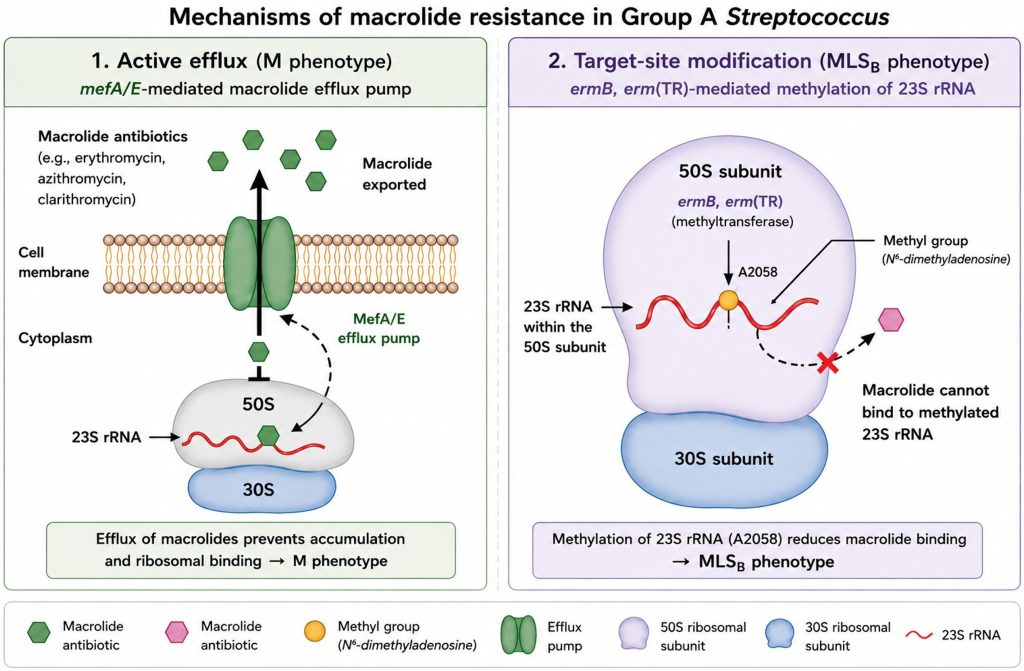
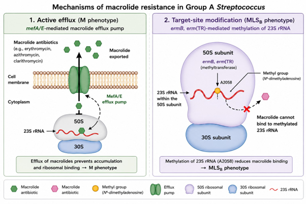
Macrolide resistance in GAS is primarily mediated by two distinct mechanisms: active drug efflux and ribosomal target site modification (Fig. 1).
The efflux mechanism is encoded by mef genes, particularly mefA and mefE, which confer the M phenotype. This mechanism results in low- to moderate-level resistance and primarily affects 14- and 15-membered macrolides, such as erythromycin and azithromycin, whereas susceptibility to lincosamides is generally preserved. In contrast, target site modifications are mediated by erm genes, including ermB, erm(T), and erm(TR). These genes encode methyltransferases that modify the 23S rRNA component of the 50S ribosomal subunit, leading to a reduced binding affinity for macrolides. This results in the macrolide-lincosamide-streptogramin B (MLSB) resistance phenotype, which confers cross-resistance to macrolides, lincosamides (e.g., clindamycin), and streptogramin B antibiotics [12,13].
Expression of erm(B) may be constitutive or inducible and is often associated with high-level MLSB resistance. In contrast, erm(TR) is more commonly associated with inducible MLSB resistance, in which clindamycin resistance may be induced during treatment. This inducible phenotype is readily detected by the disk diffusion method using the D-test, in which erythromycin and clindamycin disks are placed in close proximity. Importantly, both mef and erm genes are often located on mobile genetic elements such as transposons and prophages, facilitating horizontal gene transfer and rapid dissemination among GAS strains [5,12].
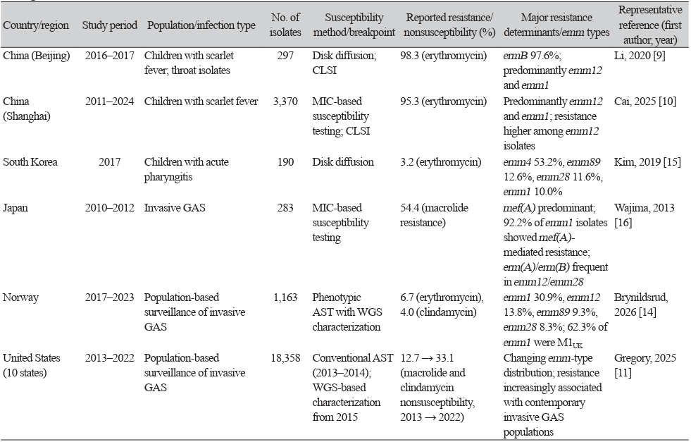
The prevalence of macrolide-resistant GAS varies substantially across geographic regions and over time, largely reflecting differences in antimicrobial consumption, clonal dissemination, and regional epidemiological characteristics [5–11] (Table 1). Macrolide resistance rates in Asia are among the highest reported worldwide. Several studies from China have reported very high macrolide resistance rates, particularly among selected pediatric and outbreak-associated populations [9,10]. Recent population-based surveillance in the United States has documented a substantial increase in macrolide and clindamycin nonsusceptibility among invasive GAS isolates, from 12.7% in 2013 to 33.1% in 2022 [11]. However, these estimates apply specifically to invasive GAS populations and should not be generalized to all GAS infections. Recent population-based genomic surveillance from Norway demonstrated relatively low erythromycin resistance among invasive GAS isolates, highlighting substantial geographic heterogeneity even among contemporary invasive-disease populations [14].
These geographic differences are influenced not only by patterns of macrolide consumption, but also by the circulation of successful resistant clones carrying mobile genetic elements. Therefore, continuous molecular surveillance is essential to monitor regional epidemiological trends and establish antimicrobial stewardship strategies.
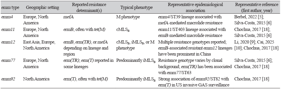
The molecular epidemiology of macrolide-resistant GAS is closely associated with the distribution of specific emm types and horizontal transfer of resistance determinants carried by mobile genetic elements [5]. emm typing, which is based on sequence variations in the emm gene encoding the M protein, has become an essential tool for investigating the epidemiology and transmission of GAS. Several emm types, including emm4, emm11, emm12, emm77, and emm92, are associated with macrolide resistance in various geographic regions [5,6,9,10,15–18]. However, associations between emm types and macrolide resistance determinants are strongly influenced by geography, study period, and clonal background [19–21] (Table 2).
Recent whole-genome sequencing (WGS) studies have further improved the understanding of GAS molecular epidemiology. In particular, the emergence of the hypervirulent M1UK lineage has attracted considerable attention because of its enhanced production of streptococcal pyrogenic exotoxin A, increased transmissibility, clonal expansion, and association with the resurgence of invasive GAS infections in several countries in Europe and United States [22–24]. Recent population-based WGS studies have further demonstrated the dynamic nature of GAS clonal epidemiology. Genomic surveillance in the United States identified frequent transmission clusters among invasive GAS isolates, with the extent of clustering varying across emm types [25]. During the post-pandemic upsurge in the United Kingdom, M1UK underwent rapid clonal expansion and international dissemination [22,24]. In contrast, recent longitudinal genomic surveillance in China identified distinct emm1 and emm12 evolutionary lineages, including the China-specific M1China lineage, and demonstrated associations between expanding sublineages and mobile genetic elements carrying antimicrobial resistance determinants [26].
The M1UK lineage provides an important example of successful clonal expansion and enhanced virulence in GAS; however, its epidemiological success should not be equated with macrolide resistance, because antimicrobial resistance varies among M1UK populations. Accordingly, genomic surveillance should evaluate virulence lineage dynamics and antimicrobial resistance determinants as related but distinct epidemiological features. These findings highlight the value of genomic surveillance for monitoring the evolution and international dissemination of clinically virulent GAS clones.
Horizontal gene transfer mediated by transposons, integrative conjugative elements, and prophages plays a critical role in the dissemination of macrolide resistance genes among GAS strains [12,13]. These mobile genetic elements facilitate the spread of erm and mef genes and contribute to the rapid emergence of resistant clones under antimicrobial selective pressure [9]. Understanding the molecular epidemiology of resistant GAS is increasingly important for identifying emerging clones, monitoring the dissemination of resistance determinants, and supporting antimicrobial stewardship and future vaccine development [27–29].
In severe invasive GAS infections, particularly toxin-mediated syndromes such as necrotizing fasciitis and streptococcal toxic shock syndrome, β-lactam therapy has traditionally been combined with a protein-synthesis inhibitor, most commonly clindamycin, to suppress toxin production [30]. Macrolides, by contrast, are primarily used as alternative agents for selected patients with β-lactam allergy [4,30]. Therefore, increased clindamycin or macrolide resistance may have important clinical implications. More recently, linezolid has emerged as a potential alternative adjunctive protein-synthesis inhibitor for invasive GAS infection. In a large retrospective cohort study using target trial emulation, adjunctive linezolid showed clinical outcomes consistent with non-inferiority to clindamycin in β-lactam-treated patients with invasive GAS infection, although prospective comparative studies are still needed [31].
Macrolide resistance may lead to microbiological treatment failure when a macrolide is used against a resistant isolate and may contribute to persistent carriage and onward transmission [30]. Furthermore, resistance mediated by erm genes frequently confers cross-resistance to clindamycin (the MLSB phenotype), potentially limiting its therapeutic value in severe invasive GAS infections. The inducible MLSB phenotype, primarily mediated by erm(TR), deserves particular clinical attention since isolates may appear susceptible to clindamycin in routine antimicrobial susceptibility testing, but become resistant during therapy. Therefore, the D-test should be routinely performed for erythromycin-resistant, clindamycin-susceptible isolates to identify inducible clindamycin resistance and guide appropriate antimicrobial therapy. Given the continuing emergence of resistant GAS clones, ongoing antimicrobial resistance surveillance and antimicrobial stewardship are essential to preserve the effectiveness of currently available therapeutic agents.
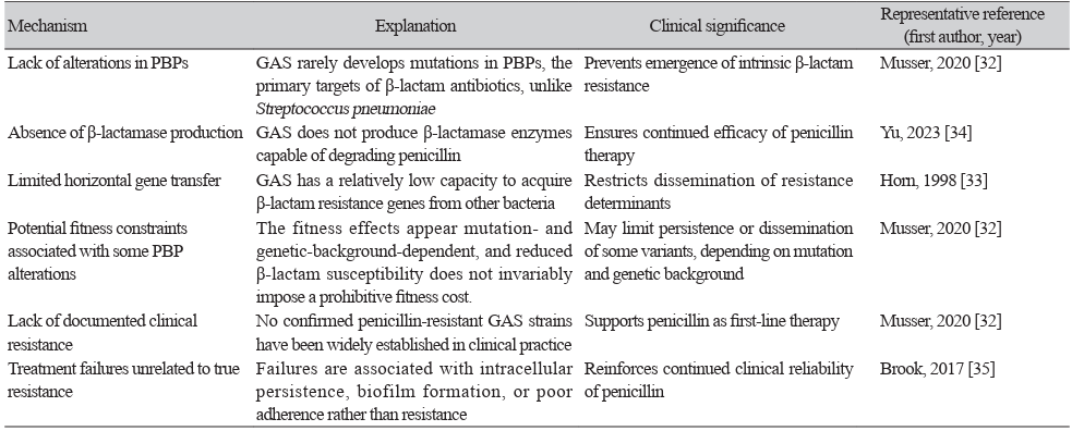
Despite more than 80 years of clinical use, GAS remains predictably susceptible to penicillin according to current clinical susceptibility criteria, and clinically established penicillin resistance has not become widespread. Nevertheless, isolates with penicillin-binding protein (PBP)2X-associated reductions in β-lactam susceptibility in vitro have been reported, warranting continued surveillance [32,33]. In contrast, other streptococcal species, particularly Streptococcus pneumoniae, have developed widespread β-lactam resistance through alterations in PBPs [5,33].
Several biological and genetic factors have been proposed to explain the sustained susceptibility of GAS to penicillin, including constraints on PBP-mediated resistance and horizontal acquisition of β-lactam resistance determinants [32–34] (Table 3). Although sporadic isolates with reduced β-lactam susceptibility have been reported, clinically established penicillin resistance in GAS remains exceedingly rare, and penicillin continues to be recommended as first-line therapy [4,35]. Continued microbiological and genomic surveillance are warranted to detect any potential emergence of reduced β-lactam susceptibility.
Although macrolide resistance remains a major concern in GAS, resistance to other antimicrobial classes has also been reported.
Fluoroquinolone resistance remains uncommon, but has occasionally been identified, particularly in countries where fluoroquinolone use is widespread. Resistance is primarily associated with mutations in the quinolone resistance-determining regions of gyrA and parC genes [16]. These mutations reduce the affinity of DNA gyrase and topoisomerase IV for fluoroquinolones. However, the overall prevalence of fluoroquinolone resistance in GAS remains low [16,18].
Tetracycline resistance is more frequently observed and is mainly mediated by the acquisition of ribosomal protection proteins encoded by tet(M) and tet(O) or less commonly by efflux pumps encoded by tet(K) and tet(L) [18,36]. These resistance determinants are frequently carried by conjugative transposons, facilitating horizontal dissemination among GAS strains.
Although resistance to quinolones and tetracyclines is currently less clinically important than macrolide resistance, continued surveillance remains important because multidrug-resistant GAS strains carrying multiple resistance determinants have occasionally been reported [37,38].
Future efforts to combat macrolide-resistant GAS should focus on continuous surveillance, antimicrobial stewardship, and development of effective preventive strategies.
Advances in molecular epidemiology, particularly WGS, have greatly improved the ability to monitor the emergence and dissemination of resistant GAS clones. The integration of genomic surveillance into national and international surveillance programs will facilitate early detection of emerging resistant lineages and improve our understanding of the evolution of GAS.
Antimicrobial stewardship programs are essential to reduce unnecessary antibiotic use and minimize the selective pressure that promotes the emergence and spread of resistant strains. Continued research on GAS vaccines offers the most promising long-term strategy for reducing the global burden of GAS infections and their sequelae. Future multidisciplinary collaborations between clinicians, microbiologists, epidemiologists, and public health authorities are needed to monitor the emergence and spread of invasive GAS lineages, improve surveillance, optimize antimicrobial use, and support vaccine implementation.
Macrolide resistance in GAS is highly heterogeneous across geographic regions, time periods, clinical populations, and circulating lineages. Both mobile resistance determinants and clonal expansion contribute to its dissemination, but reported resistance rates should be interpreted in the context of study design and infection type. β-Lactams remain the cornerstone of GAS treatment, whereas increasing macrolide and clindamycin resistance may limit alternative and adjunctive therapeutic options. Continued phenotypic and genomic surveillance using standardized methods is therefore essential for monitoring emerging resistant lineages and informing antimicrobial therapy and stewardship.
This narrative review is based solely on previously published literature and does not involve human participants, human-derived materials, or identifiable personal data. Therefore, approval from the Institutional Review Board was not required.
No potential conflicts of interest relevant to this article were reported.
This study was funded by the Ministry of Trade, Industry and Energy of Korea (RS-2024-00403563). The funders had no role in the study design, data collection and interpretation, or decision to submit the manuscript for publication.
This review article did not generate or analyze new datasets.
1. Wessels MR. Streptococcal pharyngitis. N Engl J Med 2011;364:648–55.
2. Stevens DL. Invasive group A streptococcus infections. Clin Infect Dis 1992;14:2–11.
3. Carapetis JR, Steer AC, Mulholland EK, Weber M. The global burden of group A streptococcal diseases. Lancet Infect Dis 2005;5:685–94.
4. Shulman ST, Bisno AL, Clegg HW, Gerber MA, Kaplan EL, Lee G, et al. Clinical practice guideline for the diagnosis and management of group A streptococcal pharyngitis: 2012 update by the Infectious Diseases Society of America. Clin Infect Dis 2012;55:1279–82.
5. Berbel D, González-Díaz A, López de Egea G, Càmara J, Ardanuy C. An overview of macrolide resistance in streptococci: prevalence, mobile elements and dynamics. Microorganisms 2022;10:2316.
6. Silva-Costa C, Friães A, Ramirez M, Melo-Cristino J. Macrolide-resistant Streptococcus pyogenes: prevalence and treatment strategies. Expert Rev Anti Infect Ther 2015;13:615–28.
7. Seppälä H, Klaukka T, Vuopio-Varkila J, Muotiala A, Helenius H, Lager K, et al. The effect of changes in the consumption of macrolide antibiotics on erythromycin resistance in group A streptococci in Finland. N Engl J Med 1997;337:441–6.
8. Fay K, Onukwube J, Chochua S, Schaffner W, Cieslak P, Lynfield R, et al. Patterns of antibiotic nonsusceptibility among invasive group A streptococcus infections—United States, 2006–2017. Clin Infect Dis 2021;73:1957–64.
9. Li H, Zhou L, Zhao Y, Ma L, Liu X, Hu J. Molecular epidemiology and antimicrobial resistance of group A streptococcus recovered from patients in Beijing, China. BMC Infect Dis 2020;20:507.
10. Cai J, Zhou X, Zhang C, Jiang Y, Lv P, Zhou Y, et al. Ongoing epidemic of scarlet fever in Shanghai and the emergence of M1UK lineage group A Streptococcus: a 14-year surveillance study across the COVID-19 pandemic period. Lancet Reg Health West Pac 2025;58:101576.
11. Gregory CJ, Okaro JO, Reingold A, Chai S, Herlihy R, Petit S, et al. Invasive group A streptococcal infections in 10 US States. JAMA 2025;333:1498–507.
12. Pechère JC. Macrolide resistance mechanisms in Gram-positive cocci. Int J Antimicrob Agents 2001;18(Suppl 1):S25–8.
13. Leclercq R. Mechanisms of resistance to macrolides and lincosamides: nature of the resistance elements and their clinical implications. Clin Infect Dis 2002;34:482–92.
14. Brynildsrud OB, Vollan Gjerdrum HS, Berg ES, Wester AL, Caugant DA. Evolutionary dynamics and virulence factor variability in invasive Streptococcus pyogenes in Norway, 2017–2023. mSphere 2026;11:e0077525.
15. Kim S, Lee S, Park H, Kim S. Predominance of emm4 and antibiotic resistance of Streptococcus pyogenes in acute pharyngitis in a southern region of Korea. J Med Microbiol 2019;68:1053–8.
16. Wajima T, Morozumi M, Chiba N, Shouji M, Iwata S, Sakata H, et al. Associations of macrolide and fluoroquinolone resistance with molecular typing in Streptococcus pyogenes from invasive infections, 2010–2012. Int J Antimicrob Agents 2013;42:447–9.
17. Takahashi T, Arai K, Lee DH, Koh EH, Yoshida H, Yano H, et al. Epidemiological study of erythromycin-resistant Streptococcus pyogenes from Korea and Japan by emm genotyping and multilocus sequence typing. Ann Lab Med 2016;36:9–14.
18. Chochua S, Metcalf BJ, Li Z, Rivers J, Mathis S, Jackson D, et al. Population and whole-genome sequence-based characterization of invasive group A streptococci recovered in the United States during 2015. mBio 2017;8:e01422-17.
19. Montes M, Ardanuy C, Tamayo E, Domènech A, Liñares J, Pérez-Trallero E. Epidemiological and molecular analysis of Streptococcus pyogenes isolates causing invasive disease in Spain (1998–2009): comparison with non-invasive isolates. Eur J Clin Microbiol Infect Dis 2011;30:1295–302.
20. Li Y, Rivers J, Mathis S, Li Z, Velusamy S, Nanduri SA, et al. Genomic surveillance of Streptococcus pyogenes strains causing invasive disease, United States, 2016–2017. Front Microbiol 2020;11:1547.
21. Torres RS, Torres RP, Smeesters PR, Palmeiro JK, de Messias-Reason IJ, Dalla-Costa LM. Group A Streptococcus antibiotic resistance in southern Brazil: a 17-year surveillance study. Microb Drug Resist 2011;17:313–9.
22. Lynskey NN, Jauneikaite E, Li HK, Zhi X, Turner CE, Mosavie M, et al. Emergence of dominant toxigenic M1T1 Streptococcus pyogenes clone during increased scarlet fever activity in England: a population-based molecular epidemiological study. Lancet Infect Dis 2019;19:1209–18.
23. Li Y, Nanduri SA, Van Beneden CA, Beall BW. M1UK lineage in invasive group A Streptococcus isolates from the USA. Lancet Infect Dis 2020;20:538–9.
24. Vieira A, Wan Y, Ryan Y, Li HK, Guy RL, Papangeli M, et al. Rapid expansion and international spread of M1UK in the post-pandemic UK upsurge of Streptococcus pyogenes. Nat Commun 2024;15:3916.
25. Li Y, Rivers J, Mathis S, Li Z, Chochua S, Metcalf BJ, et al. Genomic cluster formation among invasive group A streptococcal infections in the USA: a whole-genome sequencing and population-based surveillance study. Lancet Microbe 2024;5:100927.
26. You Y, Yu D, Yang C, Peng X, Xie O, Chang H, et al. Emergence of distinct Streptococcus pyogenes emm1 and emm12 lineages in China. Nat Microbiol 2026;11:1245–55.
27. Efstratiou A and Lamagni T. Epidemiology of Streptococcus pyogenes. In: Ferretti JJ, Stevens DL, et al. eds. Streptococcus pyogenes: basic biology to clinical manifestations [Internet]. 2nd ed. Oklahoma City (OK): University of Oklahoma Health Sciences Center; 2022:19. Available from: https://www.ncbi.nlm.nih.gov/books/NBK587100/
28. Dale JB and Walker MJ. Update on group A streptococcal vaccine development. Curr Opin Infect Dis 2020;33:244–50.
29. Steer AC, Law I, Matatolu L, Beall BW, Carapetis JR. Global emm type distribution of group A streptococci: systematic review and implications for vaccine development. Lancet Infect Dis 2009;9:611–6.
30. Stevens DL and Bryant AE. Severe Streptococcus pyogenes infections. 2022 Sep 7 [Updated 2022 Oct 4]. In: Ferretti JJ, Stevens DL, et al. eds. Streptococcus pyogenes: basic biology to clinical manifestations [Internet]. 2nd edition. Oklahoma City: University of Oklahoma Health Sciences Center; 2022 Oct 8. Chapter 24. Available from: https://www.ncbi.nlm.nih.gov/books/NBK587112/
31. Babiker A, Warner S, Li X, Chishti EA, Saad E, Swihart BJ, et al. Adjunctive linezolid versus clindamycin for toxin inhibition in β-lactam-treated patients with invasive group A streptococcal infections in 195 US hospitals from 2016 to 2021: a retrospective cohort study with target trial emulation. Lancet Infect Dis 2025;25:265–75.
32. Musser JM, Beres SB, Zhu L, Olsen RJ, Vuopio J, Hyyryläinen HL, et al. Reduced in vitro susceptibility of Streptococcus pyogenes to β-lactam antibiotics associated with mutations in the pbp2x gene is geographically widespread. J Clin Microbiol 2020;58:e01993-19.
33. Horn DL, Zabriskie JB, Austrian R, Cleary PP, Ferretti JJ, Fischetti VA, et al. Why have group A streptococci remained susceptible to penicillin? Report on a symposium. Clin Infect Dis 1998;26:1341–5.
34. Yu D, Guo D, Zheng Y, Yang Y. A review of penicillin binding protein and group A Streptococcus with reduced-β-lactam susceptibility. Front Cell Infect Microbiol 2023;13:1117160.
35. Brook I. Treatment challenges of group A beta-hemolytic streptococcal pharyngotonsillitis. Int Arch Otorhinolaryngol 2017;21:286–96.
36. Roberts MC. Update on acquired tetracycline resistance genes. FEMS Microbiol Lett 2005;245:195–203.
37. Centers for Disease Control and Prevention. Active bacterial core surveillance (ABCs) report, emerging infections program network, group A Streptococcus, 2023. Atlanta (GA): Centers for Disease Control and Prevention; 2025.
38. World Health Organization. Global antimicrobial resistance and use surveillance system (GLASS) report 2022. Geneva: World Health Organization; 2022.
1. Douvoyiannis M. Macrolide-resistant Mycoplasma pneumoniae in children: emphasis on the United States. Clin Pediatr (Phila) 2025;64:1169-74.

2. Antonakos N, Giamarellos-Bourboulis EJ, Niederman MS. The role of macrolides in severe community-acquired pneumonia and the potential impact of macrolide-resistant Mycoplasma pneumoniae. Curr Opin Infect Dis 2025;38:190-7.

3. Lanata MM, Wang H, Everhart K, Moore-Clingenpeel M, Ramilo O, Leber A. Macrolideresistant Mycoplasma pneumoniae infections in children, Ohio, USA. Emerg Infect Dis 2021;27:1588-97.

4. Yang S, Liu X, Han Y, Wang H, Mei Y, Wang H, et al. Clinical characteristics and associated factors of macrolide-resistant Mycoplasma pneumoniae pneumonia in children: a systematic review and meta-analysis. Eur J Clin Microbiol Infect Dis 2025;44:1505-22.

5. Ouchi K and Oishi T. Recent global trends of macrolide-resistant Mycoplasma pneumoniae: why has the macrolide-resistant rate decreased in Japan? Pediatr Infect Dis J 2025;44:e446-e9.

6. Lee JK, Lee JH, Lee H, Ahn YM, Eun BW, Cho EY, et al. Clonal expansion of macrolideresistant sequence type 3 Mycoplasma pneumoniae, South Korea. Emerg Infect Dis 2018;24:1465-71.

7. Tam KK, Suster CJE, Fong W, Golubchik T, Sivalingam V, Jeoffreys N, et al. Genomic surveillance reveals emergence and spread of macrolide-resistant Mycoplasma pneumoniae in Australia during the 2023-2024 epidemic. J Infect Dis 2026;jiag163. online ahead of print.

8. Zhu X, Tang Y, Spruyt K, Estill J, Florez ID, Amer YS, et al. Global molecular epidemiology of Mycoplasma pneumoniae and its association with macrolide resistance and disease severity: a systematic review and meta-analysis. Emerg Microbes Infect 2026;15:2614742.

9. Kim K, Jung S, Kim M, Park S, Yang HJ, Lee E. Global trends in the proportion of macrolideresistant Mycoplasma pneumoniae infections: a systematic review and meta-analysis. JAMA Netw Open 2022;5:e2220949.

10. Hong KB, Choi EH, Lee HJ, Lee SY, Cho EY, Choi JH, et al. Macrolide resistance of Mycoplasma pneumoniae, South Korea, 2000-2011. Emerg Infect Dis 2013;19:1281-4.

11. Miyashita N, Ogata M, Fukuda N, Yamura A, Ito T. Macrolide-resistant Mycoplasma pneumoniae infection prevalence increases again in Osaka. Respir Investig 2025;63:517-20.

12. Yan C, Chen Y, Ren X, Jia X, Du B, Zhao H, et al. In vitro antimicrobial susceptibility of Mycoplasma pneumoniae isolates across different regions of China in 2023. JAC Antimicrob Resist 2025;7:dlaf124.

13. Waites KB, Ratliff A, Crabb DM, Xiao L, Qin X, Selvarangan R, et al. Macrolide-resistant Mycoplasma pneumoniae in the United States as determined from a national surveillance program. J Clin Microbiol 57:10.1128/jcm.00968-19.

14. Korneenko EV, Rog IS, Chudinov IK, Kozyreva AI, Sandanova DA, Pavlenko AV, et al. Genomic surveillance of the pneumonia outbreak caused by Mycoplasma pneumoniae among patients in Russia from January 2024 to June 2025. Front Cell Infect Microbiol 2026;16:1736929.

15. Rivaya B, Jordana-Lluch E, Fernández-Rivas G, Molinos S, Campos R, Méndez-Hernández M, et al. Macrolide resistance and molecular typing of Mycoplasma pneumoniae infections during a 4 year period in Spain. J Antimicrob Chemother 2020;75:2752-9.

16. Huang C, Yang J, Cao J, Ren H, Gao N, Liu J. Antimicrobial susceptibility and molecular characteristics of Mycoplasma pneumoniae isolates from children in Xi’an regions of China. Front Pediatr 2025;13:1660687.

17. Jiang FC, Wang RF, Chen P, Dong LY, Wang X, Song Q, et al. Genotype and mutation patterns of macrolide resistance genes of Mycoplasma pneumoniae from children with pneumonia in Qingdao, China, in 2019. J Glob Antimicrob Resist 2021;27:273-8.

18. Wang N, Xu X, Xiao L, Liu Y. Novel mechanisms of macrolide resistance revealed by in vitro selection and genome analysis in Mycoplasma pneumoniae. Front Cell Infect Microbiol 2023;13:1186017.

19. Xie P, Zhang Y, Qin Y, Fang Y, Yang N, Bai Y, et al. Macrolide resistance in Mycoplasma pneumoniae in adult patients. Front Cell Infect Microbiol 2025;15:1496521.

20. Lee JK, Seong MW, Yun KW, Choi EH. Association of tandem repeat number variabilities in subunit S of the type I restriction-modification system with macrolide resistance in Mycoplasma pneumoniae. J Clin Med 2022;11:715.

21. Guiraud J, Ginevra C, Laurier-Nadalié C, Balcon C, Semanas Q, Gardette M, et al. Multiplex amplicon sequencing reveals polyclonality and low macrolide resistance in the French Mycoplasma pneumoniae outbreak, 2023-2024. Int J Infect Dis 2026;166:108506.

22. Yun KW. Growing threat of macrolide-resistant Mycoplasma pneumoniae among children: what we know and what we need. J Korean Med Sci 2025;40:e317.

23. Xie P, Liu J, Feng Y, Zhao T, Niu W, Fang Y, et al. Evolution and characteristics of tetracycline resistance in Mycoplasma pneumoniae. Microbiol Spectr 2025;13:e03398-24.

24. Wang A, Wu Z, Huang Y, Zhou H, Wu L, Jia C, et al. A 3D-printed microfluidic device for qPCR detection of macrolide-resistant mutations of Mycoplasma pneumoniae. Biosensors (Basel) 2021;11:427.

25. Dumke R. Molecular tools for typing Mycoplasma pneumoniae and mycoplasma genitalium. Front Microbiol 2022;13:904494.

26. Hong JH, Chun JK, Uh Y, Oh KJ, Kim J, Yoon KJ. Two cases of Mycoplasma pneumoniae pneumonia with A2063G mutation in the 23S rRNA gene in siblings. Ann Lab Med 2013;33:65-8.

27. Uh Y, Hong JH, Oh KJ, Cho HM, Park SD, Kim J, et al. Macrolide resistance of Mycoplasma pneumoniae and its detection rate by real-time PCR in primary and tertiary care hospitals. Ann Lab Med 2013;33:410-4.

28. Lee JK, Lee T, Kim YJ, Kim DR, Shin A, Kang HM, et al. Clinical manifestations, macrolide resistance, and treatment utilization trends of Mycoplasma pneumoniae pneumonia in children and adolescents in South Korea. Microorganisms 2024;12:1806.

29. Cho MC, Kim H, An D, Lee M, Noh SA, Kim MN, et al. Comparison of sputum and nasopharyngeal swab specimens for molecular diagnosis of Mycoplasma pneumoniae, Chlamydophila pneumoniae, and Legionella pneumophila. Ann Lab Med 2012;32:133-8.

30. Wang G, Wu P, Tang R, Zhang W. Global prevalence of resistance to macrolides in Mycoplasma pneumoniae: a systematic review and meta-analysis. J Antimicrob Chemother 2022;77:2353-63.

31. Zhao F, Liu G, Wu J, Cao B, Tao X, He L, et al. Surveillance of macrolide-resistant Mycoplasma pneumoniae in Beijing, China, from 2008 to 2012. Antimicrob Agents Chemother 2013;57:1521-3.

32. Chen W, Qi H, Jiao W, Guo Y, Wang Y, Liu J, et al. Etiological characteristics of Mycoplasma pneumoniae infection and macrolide resistance in children in Beijing: a retrospective study from 2018 to 2024. Eur J Clin Microbiol Infect Dis 2025;44:2779-91.

33. Lee E, Cho HJ, Hong SJ, Lee J, Sung H, Yu J. Prevalence and clinical manifestations of macrolide resistant Mycoplasma pneumoniae pneumonia in Korean children. Korean J Pediatr 2017;60:151-7.

34. Sung M, Roh EJ, Lee ES, Lee JY, Kim HB, Ahn Y, et al. Assessment of variables associated with prolonged admission duration in children with Mycoplasma pneumoniae pneumonia. Clin Respir J 2022;16:756-67.

35. Morozumi M, Tajima T, Sakuma M, Shouji M, Meguro H, Saito K, et al. Sequence type changes associated with decreasing macrolide-resistant Mycoplasma pneumoniae, Japan. Emerg Infect Dis 2020;26:2210-3.

36. Oishi T and Ouchi K. Recent trends in the epidemiology, diagnosis, and treatment of macrolide-resistant Mycoplasma pneumoniae. J Clin Med 2022;11:1782.

37. Wu TH, Fang YP, Liu FC, Pan HH, Yang YY, Song CS, et al. Macrolide-resistant Mycoplasma pneumoniae infections among children before and during COVID-19 pandemic, Taiwan, 2017-2023. Emerg Infect Dis 2024;30:1692-6.
38. Rothstein TE, Cunningham SA, Rieke RA, Mainella JM, Mutchler MM, Patel R. Macrolide resistance in Mycoplasma pneumoniae, Midwestern United States, 2014 to 2021. Antimicrob Agents Chemother 2022;66:e02432-21.

39. Leber AL, Embry T, Everhart K, Taveras J, Oyeniran SJ, Wang H. Macrolide-resistant Mycoplasma pneumoniae infections among children after COVID-19 pandemic, Ohio, USA. Emerg Infect Dis 2025;31:555-8.

40. Wang H, Embry T, Everhart K, Szekely M, Taveras J, Oyeniran SJ, et al. Macrolide-resistant Mycoplasma pneumoniae in Children, Ohio, USA. Emerg Infect Dis 2025;31:2341-3.

41. Chironna M, Sallustio A, Esposito S, Perulli M, Chinellato I, Di Bari C, et al. Emergence of macrolide-resistant strains during an outbreak of Mycoplasma pneumoniae infections in children. J Antimicrob Chemother 2011;66:734-7.

42. Loconsole D, De Robertis AL, Sallustio A, Centrone F, Morcavallo C, Campanella S, et al. Update on the epidemiology of macrolide-resistant Mycoplasma pneumoniae in Europe: a systematic review. Infect Dis Rep 2021;13:811-20.

43. Meyer Sauteur PM, Beeton ML, Uldum SA, Bossuyt N, Vermeulen M, Loens K, et al. Mycoplasma pneumoniae detections before and during the COVID-19 pandemic: results of a global survey, 2017 to 2021. Euro Surveill 2022;27:2100746.

44. Chen J, Wang Y, Cheng J, Ma Y, Zhang X, Bai X, et al. Synergistic impact of macrolide resistance and H3N2 infection on M. pneumoniae outbreak in children. Microbiol Spectr 2025;13:e01844-24.

45. Lee JK, Choi YY, Sohn YJ, Kim KM, Kim YK, Han MS, et al. Persistent high macrolide resistance rate and increase of macrolide-resistant ST14 strains among Mycoplasma pneumoniae in South Korea, 2019-2020. J Microbiol Immunol Infect 2022;55:910-6.

46. Wang Y, Xu B, Wu X, Yin Q, Wang Y, Li J, et al. Increased macrolide resistance rate of M3562 Mycoplasma pneumoniae correlated with macrolide usage and genotype shifting. Front Cell Infect Microbiol 2021;11:675466.

47. Jiang TT, Sun L, Wang TY, Qi H, Tang H, Wang YC, et al. The clinical significance of macrolide resistance in pediatric Mycoplasma pneumoniae infection during COVID-19 pandemic. Front Cell Infect Microbiol 2023;13:1181402.

48. Fatima Z, Jayaratne P, Arrabi A, Rutherford C, Leto D, Smieja M, et al. Macrolide resistance and P1 cytadhesin genotyping of Mycoplasma pneumoniae during outbreak, Canada, 20242025. Emerg Infect Dis 2025;31:2316-9.

49. Arfaatabar M, Sadeghi Y, Goodarzi NN, Pourmand MR. Molecular insights into macrolide resistant Mycoplasma pneumoniae isolates from outpatient clinics in Tehran, Iran. Sci Rep 2026;16:7432.