Annals of Clinical Microbiology, The official Journal of the Korean Society of Clinical Microbiology

6

Weeks in Review

4

Weeks to Publication
Indexed in KCI, KoreaMed, Synapse, DOAJ
Open Access, Peer Reviewed
pISSN 2288-0585 eISSN 2288-6850
Review article

Evolving molecular epidemiology and antimicrobial resistance of Streptococcus agalactiae in South Korea and East Asia: a narrative review

Department of Laboratory Medicine, Wonju Severance Christian Hospital, Yonsei University Wonju College of Medicine, Wonju, Korea

Ann Clin Microbiol 2026;29(3):12. https://doi.org/10.5145/ACM.2026.29.3.12
Received on 24 June 2026, Revised on 20 July 2026, Accepted on 23 July 2026, Published on 16 September 2026.
Copyright © Korean Society of Clinical Microbiology.
This is an Open Access article which is freely available under the Creative Commons Attribution-NonCommercial-NoDerivatives 4.0 International License (CC BY-NC-ND) (https://creativecommons.org/licenses/by-nc-nd/4.0/).

Abstract

Streptococcus agalactiae (group B Streptococcus, GBS) remains a major pathogen in neonatal and adult invasive diseases, while maternal colonization continues to shape prevention strategies for early-onset disease. This narrative review examines how changes in capsular serotypes, sequence types (STs), clonal complexes (CCs), and antimicrobial resistance (AMR) determinants are reshaping the molecular epidemiology of GBS, with emphasis on South Korea and comparisons across East Asia. Conventional serotyping and multilocus sequence typing remain useful for lineage definition, whereas whole-genome sequencing increasingly enables integrated assessment of serotype, clonal background, virulence factors, and resistance determinants. Major lineages show distinct epidemiologic and resistance trajectories. Serotype III/CC17 remains strongly associated with neonatal invasive disease and has acquired multidrug resistance in some settings through mobile genetic elements. In South Korea, serotype III/CC19 has shifted from ST19 toward ST335, accompanied by increasing fluoroquinolone resistance driven by alterations in gyrA and parC. Serotype Ib/CC12 has emerged as a high-risk lineage in parts of East Asia, although resistance phenotypes vary geographically and differ substantially across STs or populations. Serotype VIII/CC1, particularly ST2, has expanded in South Korea while generally retaining lower resistance to several non-beta-lactam agents. Across Korean studies, apparent temporal AMR changes warrant caution because they may reflect heterogeneity in population composition, sample collection and laboratory examination. Continued genomic surveillance with standardized susceptibility testing and comparable sampling helps distinguish true clonal evolution from surveillance artifacts. The resulting evidence landscape should inform intrapartum prophylaxis, empirical therapy, and antimicrobial stewardship for neonates, pregnant women, and adults with invasive GBS disease.

Keywords

Drug resistance, bacterial; Molecular epidemiology; Multilocus sequence typing; Streptococcus agalactiae; Whole genome sequencing

Introduction

Streptococcus agalactiae is a facultative anaerobic, catalase-negative pathogen that appears as Gram-positive diplococci or chains. In 1884, French veterinarians and microbiologists Edmond Nocard and Henri Mollereau isolated the causative agent from the mammary tissue of cows during a mastitis outbreak and named it “Streptococcus de la mammite” [1]. In 1896, Karl Bernhard Lehmann and Rudolf Neumann proposed the scientific name Streptococcus agalactiae, combining the Greek roots “a-” (absence) and “galaktos” (milk) to signify the absence of milk secretion [2]. In 1933, Rebecca Lancefield analyzed serological differences in the cell wall carbohydrate antigen “substance C” using rabbit immune sera, classifying S. agalactiae as Group B and establishing the clinical term “Group B Streptococcus (GBS)” [3]. Beyond humans and cattle, GBS infects a broad range of hosts, highlighting the importance of monitoring its antimicrobial resistance (AMR) from a One Health perspective [4]. Although clinical GBS isolates show increasing and diverse antimicrobial resistance worldwide [5], studies have not yet systematically defined how resistance patterns vary by GBS strain type or patient age. This narrative review summarizes the global molecular epidemiology of GBS while focusing on recent changes in clonal distribution and antimicrobial resistance in South Korea, with comparisons to other East Asian countries.

Literature search and selection

We searched PubMed and Google Scholar from March 12 to June 17, 2026 using combinations of “Streptococcus agalactiae” or “group B Streptococcus” with “molecular epidemiology,” “serotype,” “sequence type,” “clonal complex,” “whole-genome sequencing,” “antimicrobial resistance,” “South Korea,” and “East Asia.” We prioritized peer-reviewed human studies reporting epidemiologic, antimicrobial susceptibility, or genomic data and retained seminal older studies when required for historical or mechanistic context. Reference lists of relevant articles were additionally screened for eligible studies. Because this was a narrative review, study selection was intended to provide a representative rather than exhaustive synthesis. We limited the review to academic literature published in English or Korean. We included studies that provided relevant clinical data but excluded studies when we could not determine sufficient information about the patient population. The first author conducted the literature review and prepared the initial manuscript. The corresponding author verified the accuracy of the cited references and their supporting statements and revised the manuscript to ensure a coherent overall structure and logical flow.

Global epidemiology

GBS colonizes the intestinal and vaginal microbiomes of women, establishing intermittent, transient, or persistent rectovaginal colonization [6]. Host immunological vulnerability, combined with environmental factors, transforms this commensal bacterium into a pathogen that causes life-threatening invasive disease [7]. Clinically, GBS infections fall into two distinct epidemiological categories: invasive infections in infants and invasive infections in adults. Neonatal infections are categorized into two groups based on the time of onset after birth. Early-onset disease (EOD) is defined as a GBS infection occurring within 0–6 days after birth, whereas late-onset disease (LOD) is defined as an invasive infection occurring between 7 and 89 days after birth [8]. In non-pregnant adults, GBS acts as an opportunistic pathogen that primarily affects older adults with immunosenescence or comorbidities such as diabetes, malignancies, chronic kidney disease, and cardiovascular disease [9]. Following the nationwide implementation of prenatal screening and intrapartum antibiotic prophylaxis (IAP), the United States reduced the EOD rate by more than 80%, from 1.7 cases per 1,000 live births in the early 1990s to 0.17 in 2022 and 0.18 in 2023 [10]. Conversely, the incidence of invasive GBS infections in adults increased significantly, from 8.1 cases per 100,000 individuals in 2008 to 10.9 cases per 100,000 individuals in 2016 (P = 0.002) [11]. Reflecting this trend, the incidence of invasive GBS infections among non-pregnant adults in Belgium more than doubled, from 3.7 to 8.2 cases per 100,000 individuals between 2009 and 2018 [12]. Japan maintains guidelines for universal prenatal GBS screening and IAP, achieving a very low EOD incidence of 0.10 cases per 1,000 live births [13]. China has an incidence of invasive GBS disease among infants under 3 months, reaching 0.31 cases per 1,000 live births [14]. In Southeast Asia, improper preparation of farmed freshwater fish causes GBS infections, even in young, healthy adults without comorbidities [15,16]. In South Korea, a multicenter retrospective study conducted at 14 university hospitals between 1996 and 2005 revealed that EOD accounted for only 20.4% of cases, whereas LOD accounted for the remaining 79.6% [17]. Maternal GBS colonization rates in South Korea increased from 8.0% in 2008 to 19.8% in 2019 [18,19]. Between 2006 and 2015, 93.2% of 74 adult patients with invasive GBS infections in South Korea had at least one severe chronic comorbidity [20]. Collectively, these findings indicate that GBS epidemiology varies according to host population, prevention policies, geographic setting, exposure patterns, and local clonal composition.

Molecular typing

Molecular epidemiological characterization, particularly serotype and clonal complex (CC) analyses, elucidates the genetic background that determines GBS disease patterns and tissue invasiveness [21]. Capsular polysaccharide diversity defines 10 distinct GBS serotypes (Ia, Ib, II, and III–IX), and their prevalence and distribution are influenced by geographic region, ethnicity, and clinical manifestation [22]. Historically, investigators have used phenotypic assays, such as latex agglutination and enzyme-based immunoassays, to classify GBS serotypes [23,24]. More recently, genetic analysis of the cps gene locus has provided a reliable method for serotype classification [25,26]. Multilocus sequence typing, based on seven housekeeping genes, enables precise strain characterization without ambiguity in GBS epidemiological studies. By grouping unique sequence types (STs) using phylogenetic and clustering algorithms, this method delineates the GBS population structure into broader CCs [27]. In this hierarchical framework, CCs represent the overarching genetic lineages composed of closely related clones, whereas STs represent the individual STs within each complex [27]. For instance, CC17 includes ST17 as its predominant ST, defining a relatively homogeneous, hypervirulent lineage that is strongly associated with invasive neonatal infections and capsular serotype III [27].

Because horizontal gene transfer and clonal expansion drive the dissemination of AMR, combining traditional phenotypic susceptibility testing with nucleic acid amplification tests enhances resistance surveillance and facilitates rapid diagnosis [28]. GBS harbors diverse resistance genes that mediate target modification (pbp, erm, gyr, and par), target protection (tet, msr, and lsa), enzymatic inactivation (lnu and cat), or active efflux (mef) [2934]. Although conventional PCR-based assays are limited to detecting specific target genes, whole-genome sequencing (WGS) enables comprehensive molecular characterization by simultaneously analyzing multiple genetic features [35]. In GBS isolates from South Korean women of childbearing age around the year 2000, the prevalence of tetM in non-multidrug-resistant (non-MDR) strains significantly decreased from 64.9% to 22.5% (P < 0.001), whereas the prevalence of tetO in MDR strains significantly increased from 0% to 12.0% (P = 0.019). Notably, both non-MDR and MDR strains demonstrated substantial increases in levofloxacin resistance associated with gyrA and parC mutations [36].

Hypervirulent CC17 serotype III GBS

Strains belonging to serotype III and ST17 constitute a hypervirulent GBS clone responsible for more than 60% of neonatal meningitis and LOD cases in many surveillance studies [37]. The hypervirulent GBS adhesin gene (hvgA) and the surface-anchored protein gene srr2 contribute to this high virulence by promoting blood-brain barrier penetration and meningeal invasion [38]. Recent studies have confirmed that historically susceptible ST17 serotype III clones have evolved into highly MDR strains through horizontal gene transfer [39]. These strains acquired a large (~75-kb) integrative and conjugative element, ICESag37, which stably integrated ermB and tetO into the genome while replacing the pilus island 1 gene cluster and the insertion sequence ISSag5 [37]. Colonizing CC17 GBS isolates from South Korean women of childbearing age belonged exclusively to serotype III. Before the 2000s, these strains exhibited resistance only to tetracycline; subsequently, they acquired resistance to erythromycin and clindamycin [36].

Predominant CC19 serotype III GBS

Serotype III represents the most prevalent serotype among clinical GBS isolates in South Korea, with ST19 being the most common ST within this serotype [40,41]. Among colonizing CC19 serotype III GBS isolates from women of childbearing age, the proportion of ST19 decreased from 85.7% (12/14) during 1994–2000 to 30.6% (11/36) during 2017–2022, whereas ST335 emerged as the predominant ST, accounting for 50.0% (18/36) of isolates [36]. The overall proportion of colonizing CC19 serotype III GBS isolates among South Korean women of childbearing age significantly decreased from a peak of 37.8% during 1994–2000 to 17.2% during 2017–2022 (P = 0.004) [36]. Although ST19 serotype III strains maintained resistance to erythromycin, clindamycin, tetracycline, and chloramphenicol, their levofloxacin resistance rate significantly increased from 0% to 63.6% [36]. AMR profiles of this GBS lineage vary across East Asian countries and among specific STs. From September to December 2011, ST19 serotype III accounted for 80.0% of clinically isolated levofloxacin-resistant GBS strains across eight Chinese cities [42]. In contrast, prenatal screening conducted between 2017 and 2021 identified 12 CC19 serotype III isolates (10 ST335 and 2 ST27) that exhibited no levofloxacin resistance in Japan [43]. In South Korea, clinical ST19 serotype III GBS strains isolated between 2010 and 2011 carried lnuB and lsaE, displaying a unique phenotype of clindamycin resistance combined with erythromycin susceptibility [44].

Emergent CC12 serotype Ib GBS

Although historical data have identified ST17 serotype III as the primary hypervirulent lineage causing neonatal invasive disease, the ST12/CC12 serotype Ib lineage has rapidly emerged across East and Southeast Asia as a high-risk lineage [45,46]. In Taiwan, CC12 serotype Ib strains accounted for only 7.4% of neonatal invasive isolates collected between 2003 and 2020, yet caused complicated sepsis in 71.4% of cases and were associated with a sepsis-related mortality rate of 42.9%, as well as 100% resistance to erythromycin and clindamycin [45]. Among the 15 CC12 serotype Ib GBS isolates collected from pediatric patients at a university hospital in Shanghai between 2009 and 2020, 13 (86.7%) were associated with bloodstream infections, and the mortality rate among these 13 patients was 30.8% (4/13) [46]. In South Korea, the proportion of colonizing CC12 serotype Ib GBS isolates among women of childbearing age significantly declined from 27.0% during 1994–2000 to 13.4% during 2017–2022 (P = 0.034) [36]. However, the single ST12 serotype Ib isolate identified during 1994–2000 exhibited resistance only to tetracycline, whereas all three isolates identified during 2017–2022 were MDR, exhibiting resistance to erythromycin, clindamycin, and tetracycline [36].

Pervasive CC1 serotype VIII GBS

Serotype VIII GBS gained clinical attention after it was identified in 35.6% (26/73) of vaginal GBS isolates from pregnant women in Japan between 1992 and 1994 [47]. South Korea mirrors this trend, with serotype VIII accounting for 20.0% of colonizing GBS isolates from pregnant women between 2017 and 2019 [19]. The proportion of serotype VIII among colonizing GBS isolates from women of childbearing age significantly increased from 2.7% during 1994–2000 to 15.3% during 2017–2022 (P = 0.037), with the predominant ST within CC1 shifting from ST1 to ST2 [36]. Although ST2 accounted for 87.8% (36/41) of sepsis cases caused by serotype VIII GBS, these strains maintained low resistance rates (below 20.0%) to erythromycin, clindamycin, and levofloxacin [48]. Colonizing serotype VIII/ST2 GBS isolates from women of childbearing age consistently exhibited a similarly low resistance profile [36].

Shifting AMR profiles

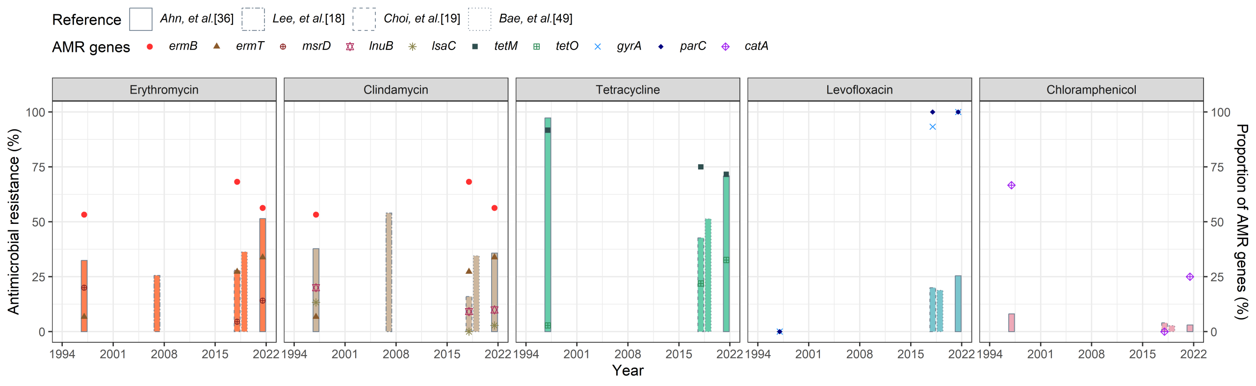
GBS causes a wide spectrum of clinical infections. Maternal colonization is a major focus of prevention because of its association with neonatal EOD, while invasive GBS disease among non-pregnant and older adults represents an increasingly important clinical burden. In South Korea, longitudinal surveillance of AMR profiles among these colonizing isolates has revealed dynamic temporal changes [36]. Specifically, tetracycline resistance has recently shown a slight decline after remaining at consistently high levels, whereas erythromycin resistance has continued to fluctuate. Clindamycin resistance, which had previously shown a declining trend, has recently resurged [18,19,36,49]. Recent advances in WGS have enabled comprehensive investigations into the correlation between resistance phenotypes and their underlying genotypic profiles, revealing substantial evolutionary shifts in resistance determinants among South Korean GBS strains [35,36]. Genotypically, the prevalence of the tetracycline resistance gene tetM has decreased in South Korean studies, whereas that of tetO has increased sharply. Similarly, although the macrolide-lincosamide-streptogramin B resistance gene ermB showed no significant change, the prevalence of ermT increased [36]. In clinical practice, tetracycline is contraindicated during pregnancy, and penicillin-class antibiotics remain the first-line agents for IAP. For GBS-positive patients with a high risk of anaphylaxis to penicillin, clindamycin is an option only when the isolate is susceptible; susceptibility testing is therefore essential for selecting appropriate IAP [6,50]. Therefore, the recent increase in ermT-mediated clindamycin resistance among South Korean isolates warrants close clinical vigilance. Furthermore, the increasing colonization of levofloxacin-resistant GBS in South Korea underscores a concerning trend in which resistant strains may be selected under antimicrobial pressure [36]. Antimicrobial exposure may contribute to the selection and expansion of resistant GBS lineages; however, current surveillance data do not establish that antimicrobial selection caused the observed increase in GBS colonization. Changes in screening practices, specimen collection, culture methods, referral patterns, and population characteristics may also have contributed. In addition, advances in diagnostic technologies may have contributed to the observed increase in GBS colonization among South Korean women. Across Korean studies, AMR estimates varied substantially according to the study population and laboratory methodology (Fig. 1). Despite this heterogeneity, fluoroquinolone resistance and gyrA/parC alterations appear more prominent in recent datasets, whereas tetracycline- and macrolide/lincosamide-resistance determinants show lineage-specific redistribution. These cross-study comparisons should be interpreted descriptively rather than as a continuous national time trend.


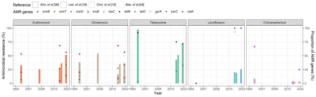
Fig. 1. Antimicrobial resistance (AMR) profiles and associated genes of group B Streptococcus (GBS) isolated from South Korean women of childbearing age and pregnant women. The x-axis shows the reference studies included in the analysis and the corresponding bacterial isolate collection periods. Each column uses a distinct border to distinguish individual studies and is positioned at the midpoint of the isolate collection period for that study. Column heights represent phenotypic antimicrobial resistance rates (left y-axis). Distinct geometric symbols represent the proportions of isolates carrying AMR genes or resistance-associated mutations among GBS isolates resistant to the corresponding antimicrobial agent in each study (right y-axis). Estimates were derived from independent studies with heterogeneous populations, sampling strategies, and laboratory methods and therefore should not be interpreted as a continuous longitudinal national trend.

Conclusion

Selective pressures and horizontal gene transfer may contribute to the dissemination of MDR across distinct GBS lineages, posing a major challenge to standard clinical management. The genomic integration of mobile genetic elements, such as ICESag37 in hypervirulent CC17 serotype III clones, incorporates the ermB and tetO genes, stabilizing resistance phenotypes. Recent epidemiological data from South Korea revealed a dramatic increase in fluoroquinolone resistance. The predominant CC19 serotype III lineage showed a clear genotypic shift from ST19 to ST335, accompanied by a sharp increase in levofloxacin resistance mediated by gyrA and parC mutations. Serotype Ib/CC12 has emerged as a clinically important lineage in East Asia, but its AMR phenotype varies geographically and among STs. Taiwanese neonatal ST12/CC12 isolates showed uniformly high resistance to erythromycin and clindamycin [45], whereas fluoroquinolone resistance reported in South Korean adult serotype Ib isolates involved other STs [20]. The rapidly expanding serotype VIII/ST2/CC1 clone among South Korean adults maintains comparatively lower resistance rates to several non-beta-lactam agents, distinguishing it from other MDR lineages. These shifting AMR profiles and the emergence of specific MDR clones threaten the efficacy of empirical therapy. Effective mitigation of this evolving resistance landscape requires ongoing molecular surveillance and targeted antibiotic stewardship.

Ethics statement

This study did not involve human participants. Therefore, institutional review board (IRB) approval and informed consent were not required.

Conflicts of interest

Young Uh has been a Statistical editor of the Annals of Clinical Microbiology since January 2024. However, he was not involved in the review process of this article. No other potential conflicts of interest relevant to this article were reported.

Funding

None.

Data availability

This review article did not generate or analyze new datasets.

References

1. Nocard E and Mollereau H. Sur une mammite contagieuse des vaches laitieres. Bull Acad Vet Fr 1884;38:308-14.

2. Skerman VBD, McGowan V, Sneath PHA. Approved lists of bacterial names. Int J Syst Evol Microbiol 1980;30:225-420.

3. Lancefield RC. A serological differentiation of human and other groups of hemolytic streptococci. J Exp Med 1933;57:571-95.

4. Oliveira LMA, Simões LC, Costa NS, Zadoks RN, Pinto TCA. The landscape of antimicrobial resistance in the neonatal and multi-host pathogen group B Streptococcus: review from a One Health perspective. Front Microbiol 2022;13:943413.

5. Hsu CY, Moradkasani S, Suliman M, Uthirapathy S, Zwamel AH, Hjazi A, et al. Global patterns of antibiotic resistance in group B Streptococcus: a systematic review and meta-analysis. Front Microbiol 2025;16:1541524.

6. American College of Obstetricians and Gynecologists. Prevention of group B streptococcal early-onset disease in newborns: ACOG Committee Opinion, Number 797. Obstet Gynecol 2020;135:e51-e72.

7. Shabayek S and Spellerberg B. Group B streptococcal colonization, molecular characteristics, and epidemiology. Front Microbiol 2018;9:437.

8. Coggins SA and Puopolo KM. Neonatal group B Streptococcus disease. Pediatr Rev 2024;45:63-73.

9. Ali M, Razok A, Rehman A, Al-Wali W, Al Maslamani M, Hadi HA. Descriptive review: global spectrum of invasive group B streptococcal disease in nonpregnant adults: epidemiology, risk factors and clinical presentations. Eur J Clin Microbiol Infect Dis 2026. Epub ahead of print. https://doi.org/10.1007/s10096-026-05543-z

10. Centers for Disease Control and Prevention. Group B strep surveillance and trends. https://www.cdc.gov/group-b-strep/php/surveillance/index.html [Online] (last visited on 2 August 2026).

11. Francois Watkins LK, McGee L, Schrag SJ, Beall B, Jain JH, Pondo T, et al. Epidemiology of invasive group B streptococcal infections among nonpregnant adults in the United States, 2008-2016. JAMA Intern Med 2019;179:479-88.

12. Graux E, Hites M, Martiny D, Maillart E, Delforge M, Melin P, et al. Invasive group B Streptococcus among non-pregnant adults in Brussels-Capital Region, 2005-2019. Eur J Clin Microbiol Infect Dis 2021;40:515-23.

13. Matsubara K and Shibata M. Group B streptococcal disease in infants in Japan. Pediatr Infect Dis J 2024;43:e3-e10.

14. Ji W, Liu H, Madhi SA, Cunnington M, Zhang Z, Dangor Z, et al. Clinical and molecular epidemiology of invasive group B Streptococcus disease among infants, China. Emerg Infect Dis 2019;25:2021-30.

15. Aiewsakun P, Ruangchai W, Thawornwattana Y, Jaemsai B, Mahasirimongkol S, Homkaew A, et al. Genomic epidemiology of Streptococcus agalactiae ST283 in Southeast Asia. Sci Rep 2022;12:4185.

16. Li C, Tse H, Zhu C, Choi GKY, Lee ALH, Yang J, et al. Invasive group B Streptococcus infections caused by hypervirulent clone of S. agalactiae sequence type 283, Hong Kong, China, 2021. Emerg Infect Dis 2025;31:149-54.

17. Park KH, Kim KH, Kang JH, Kim KN, Kim DS, Kim YK, et al. Current status and clinical presentations of invasive neonatal group B streptococcal infections in Korea. Pediatr Int 2011;53:236-9.

18. Lee BK, Song YR, Kim MY, Yang JH, Shin JH, Seo YS, et al. Epidemiology of group B streptococcus in Korean pregnant women. Epidemiol Infect 2010;138:292-8.

19. Choi SJ, Kang J, Uh Y. Recent epidemiological changes in group B Streptococcus among pregnant Korean women. Ann Lab Med 2021;41:380-5.

20. Lee H, Kim ES, Song KH, Kim HB, Park JS, Park KU. Clinical and molecular epidemiology of invasive group B Streptococcus infections in adults in a referral center in Korea. Eur J Clin Microbiol Infect Dis 2022;41:1407-13.

21. Chaguza C, Jamorzy D, Bijlsma MW, Kuijpers TW, van de Beek D, van der Ende A, et al. Population genomics of group B Streptococcus reveals the genetics of neonatal disease onset and meningeal invasion. Nat Commun 2022;13:4215.

22. Schuchat A. Epidemiology of group B streptococcal disease in the United States: shifting paradigms. Clin Microbiol Rev 1998;11:497-513.

23. Arakere G, Flores AE, Ferrieri P, Frasch CE. Inhibition enzyme-linked immunosorbent assay for serotyping of group B streptococcal isolates. J Clin Microbiol 1999;37:2564-7.

24. Slotved HC, Elliott J, Thompson T, Konradsen HB. Latex assay for serotyping of group B Streptococcus isolates. J Clin Microbiol 2003;41:4445-7.

25. Lee Y, Bae HG, Won D, Yun W, Lee H, Choi JR, et al. Comparative analysis of the molecular characteristics of group B Streptococcus isolates collected from pregnant Korean women using whole-genome sequencing. Ann Lab Med 2023;43:180-6.

26. Kapatai G, Patel D, Efstratiou A, Chalker VJ. Comparison of molecular serotyping approaches of Streptococcus agalactiae from genomic sequences. BMC Genomics 2017;18:429.

27. Jones N, Bohnsack JF, Takahashi S, Oliver KA, Chan MS, Kunst F, et al. Multilocus sequence typing system for group B streptococcus. J Clin Microbiol 2003;41:2530-6.

28. Fluit AC, Visser MR, Schmitz FJ. Molecular detection of antimicrobial resistance. Clin Microbiol Rev 2001;14:836-71.

29. Piccinelli G, Carlentini G, Gargiulo F, Caruso A, De Francesco MA. Analysis of point mutations in the pbp2x, pbp2b, and pbp1a genes of Streptococcus agalactiae and their relation with a reduced susceptibility to cephalosporins. Microb Drug Resist 2017;23:1019-24.

30. Li W, Atkinson GC, Thakor NS, Allas U, Lu CC, Chan KY, et al. Mechanism of tetracycline resistance by ribosomal protection protein Tet(O). Nat Commun 2013;4:1477.

31. Leclercq R. Mechanisms of resistance to macrolides and lincosamides: nature of the resistance elements and their clinical implications. Clin Infect Dis 2002;34:482-92.

32. Hayes K, Cotter L, Barry L, O’Halloran F. Emergence of the L phenotype in group B streptococci in the south of Ireland. Epidemiol Infect 2017;145:3535-42.

33. Pozzi G and Guild WR. Two genes for chloramphenicol resistance common to staphylococci and streptococci. Eur J Epidemiol 1988;4:20-4.

34. Wehbeh W, Rojas-Diaz R, Li X, Mariano N, Grenner L, Segal-Maurer S, et al. Fluoroquinolone-resistant Streptococcus agalactiae: epidemiology and mechanism of resistance. Antimicrob Agents Chemother 2005;49:2495-7.

35. Metcalf BJ, Chochua S, Gertz RE Jr, Hawkins PA, Ricaldi J, Li Z, et al. Short-read whole genome sequencing for determination of antimicrobial resistance mechanisms and capsular serotypes of current invasive Streptococcus agalactiae recovered in the USA. Clin Microbiol Infect 2017;23:574.e7-e14.

36. Ahn K, Choi SJ, Bae HG, Lee H, Choi JR, Uh Y, et al. Increasing antimicrobial resistance profile diversity of colonizing group B Streptococcus in reproductive-aged women in Korea. Ann Lab Med 2025. Epub ahead of print. https://doi.org/10.3343/alm.2025.0439

37. Hsu JF, Tsai MH, Lin LC, Chu SM, Lai MY, Huang HR, et al. Genomic characterization of serotype III/ST-17 group B Streptococcus strains with antimicrobial resistance using whole genome sequencing. Biomedicines 2021;9:1477.

38. Jin Z, Li J, Zhou H, Wang Z, Yi L, Liu N, et al. Serotype distribution, virulence determinants and antimicrobial susceptibility of Streptococcus agalactiae isolated from young infants. Pathogens 2022;11:1355.

39. Teatero S, Ramoutar E, McGeer A, Li A, Melano RG, Wasserscheid J, et al. Clonal complex 17 group B Streptococcus strains causing invasive disease in neonates and adults originate from the same genetic pool. Sci Rep 2016;6:20047.

40. Lee JH, Cho HK, Kim KH, Lee H, Jo DS, Kim HW. Genotypic distribution of alpha-like proteins in group B Streptococcus strains isolated in Korea: implications for vaccine coverage. Infect Chemother 2025;57:218-29.

41. Choi JH, Kim TH, Kim ET, Kim YR, Lee H. Molecular epidemiology and virulence factors of group B Streptococcus in South Korea according to the invasiveness. BMC Infect Dis 2024;24:740.

42. Wang H, Zhao C, He W, Zhang F, Zhang L, Cao B, et al. High prevalence of fluoroquinolone-resistant group B streptococci among clinical isolates in China and predominance of sequence type 19 with serotype III. Antimicrob Agents Chemother 2013;57:1538-41.

43. Kawaguchiya M, Urushibara N, Aung MS, Shimada S, Nakamura M, Ito M, et al. Molecular characterization and antimicrobial resistance of Streptococcus agalactiae isolated from pregnant women in Japan, 2017-2021. IJID Reg 2022;4:143-5.

44. Takahashi T, Maeda T, Lee S, Lee DH, Kim S. Clonal distribution of clindamycin-resistant erythromycin-susceptible (CRES) Streptococcus agalactiae in Korea based on whole genome sequences. Ann Lab Med 2020;40:370-81.

45. Hsu JF, Chen YN, Chu SM, Lee WJ, Huang HR, Chiang MC, et al. Clonal complex 12 serotype Ib Streptococcus agalactiae strain causing complicated sepsis in neonates: clinical features and genetic characteristics. Microbiol Spectr 2023;11:e03778-22.

46. Liu J, Chen F, Guan H, Yu J, Yu J, Zhao J, et al. Emerging fatal Ib/CC12 hypervirulent multiresistant Streptococcus agalactiae in young infants with bloodstream infection in China. Front Microbiol 2021;12:767803.

47. Lachenauer CS, Kasper DL, Shimada J, Ichiman Y, Ohtsuka H, Kaku M, et al. Serotypes VI and VIII predominate among group B streptococci isolated from pregnant Japanese women. J Infect Dis 1999;179:1030-3.

48. Kim M, Choi SM, Ji S, Cho D, Han D, Lee A, et al. Serotype distribution and clinical characteristics of group B Streptococcus bacteremia in nonpregnant adults: a 15-year multicenter study in Korea. BMC Infect Dis 2024;24:1441.

49. Bae HG, Hong J, Kim YJ, Lee KR, Lee K, Choi SJ, et al. A retrospective national study on colonization rate and antimicrobial susceptibility of Streptococcus agalactiae in pregnant Korean women, 2018-2020. Yonsei Med J 2022;63:717-23.

50. Shin A, Kim DR, Sung JH, Yang J, Choi SJ, Roh CR, et al. Group B Streptococcus detection rate and clindamycin resistance among reproductive-age women in Korea during 2003-2022. J Korean Med Sci 2025;40:e29.

Figure 1

1. Nocard E and Mollereau H. Sur une mammite contagieuse des vaches laitieres. Bull Acad Vet Fr 1884;38:308-14.

2. Skerman VBD, McGowan V, Sneath PHA. Approved lists of bacterial names. Int J Syst Evol Microbiol 1980;30:225-420.

3. Lancefield RC. A serological differentiation of human and other groups of hemolytic streptococci. J Exp Med 1933;57:571-95.

4. Oliveira LMA, Simões LC, Costa NS, Zadoks RN, Pinto TCA. The landscape of antimicrobial resistance in the neonatal and multi-host pathogen group B Streptococcus: review from a One Health perspective. Front Microbiol 2022;13:943413.

5. Hsu CY, Moradkasani S, Suliman M, Uthirapathy S, Zwamel AH, Hjazi A, et al. Global patterns of antibiotic resistance in group B Streptococcus: a systematic review and meta-analysis. Front Microbiol 2025;16:1541524.

6. American College of Obstetricians and Gynecologists. Prevention of group B streptococcal early-onset disease in newborns: ACOG Committee Opinion, Number 797. Obstet Gynecol 2020;135:e51-e72.

7. Shabayek S and Spellerberg B. Group B streptococcal colonization, molecular characteristics, and epidemiology. Front Microbiol 2018;9:437.

8. Coggins SA and Puopolo KM. Neonatal group B Streptococcus disease. Pediatr Rev 2024;45:63-73.

9. Ali M, Razok A, Rehman A, Al-Wali W, Al Maslamani M, Hadi HA. Descriptive review: global spectrum of invasive group B streptococcal disease in nonpregnant adults: epidemiology, risk factors and clinical presentations. Eur J Clin Microbiol Infect Dis 2026. Epub ahead of print. https://doi.org/10.1007/s10096-026-05543-z

10. Centers for Disease Control and Prevention. Group B strep surveillance and trends. https://www.cdc.gov/group-b-strep/php/surveillance/index.html [Online] (last visited on 2 August 2026).

11. Francois Watkins LK, McGee L, Schrag SJ, Beall B, Jain JH, Pondo T, et al. Epidemiology of invasive group B streptococcal infections among nonpregnant adults in the United States, 2008-2016. JAMA Intern Med 2019;179:479-88.

12. Graux E, Hites M, Martiny D, Maillart E, Delforge M, Melin P, et al. Invasive group B Streptococcus among non-pregnant adults in Brussels-Capital Region, 2005-2019. Eur J Clin Microbiol Infect Dis 2021;40:515-23.

13. Matsubara K and Shibata M. Group B streptococcal disease in infants in Japan. Pediatr Infect Dis J 2024;43:e3-e10.

14. Ji W, Liu H, Madhi SA, Cunnington M, Zhang Z, Dangor Z, et al. Clinical and molecular epidemiology of invasive group B Streptococcus disease among infants, China. Emerg Infect Dis 2019;25:2021-30.

15. Aiewsakun P, Ruangchai W, Thawornwattana Y, Jaemsai B, Mahasirimongkol S, Homkaew A, et al. Genomic epidemiology of Streptococcus agalactiae ST283 in Southeast Asia. Sci Rep 2022;12:4185.

16. Li C, Tse H, Zhu C, Choi GKY, Lee ALH, Yang J, et al. Invasive group B Streptococcus infections caused by hypervirulent clone of S. agalactiae sequence type 283, Hong Kong, China, 2021. Emerg Infect Dis 2025;31:149-54.

17. Park KH, Kim KH, Kang JH, Kim KN, Kim DS, Kim YK, et al. Current status and clinical presentations of invasive neonatal group B streptococcal infections in Korea. Pediatr Int 2011;53:236-9.

18. Lee BK, Song YR, Kim MY, Yang JH, Shin JH, Seo YS, et al. Epidemiology of group B streptococcus in Korean pregnant women. Epidemiol Infect 2010;138:292-8.

19. Choi SJ, Kang J, Uh Y. Recent epidemiological changes in group B Streptococcus among pregnant Korean women. Ann Lab Med 2021;41:380-5.

20. Lee H, Kim ES, Song KH, Kim HB, Park JS, Park KU. Clinical and molecular epidemiology of invasive group B Streptococcus infections in adults in a referral center in Korea. Eur J Clin Microbiol Infect Dis 2022;41:1407-13.

21. Chaguza C, Jamorzy D, Bijlsma MW, Kuijpers TW, van de Beek D, van der Ende A, et al. Population genomics of group B Streptococcus reveals the genetics of neonatal disease onset and meningeal invasion. Nat Commun 2022;13:4215.

22. Schuchat A. Epidemiology of group B streptococcal disease in the United States: shifting paradigms. Clin Microbiol Rev 1998;11:497-513.

23. Arakere G, Flores AE, Ferrieri P, Frasch CE. Inhibition enzyme-linked immunosorbent assay for serotyping of group B streptococcal isolates. J Clin Microbiol 1999;37:2564-7.

24. Slotved HC, Elliott J, Thompson T, Konradsen HB. Latex assay for serotyping of group B Streptococcus isolates. J Clin Microbiol 2003;41:4445-7.

25. Lee Y, Bae HG, Won D, Yun W, Lee H, Choi JR, et al. Comparative analysis of the molecular characteristics of group B Streptococcus isolates collected from pregnant Korean women using whole-genome sequencing. Ann Lab Med 2023;43:180-6.

26. Kapatai G, Patel D, Efstratiou A, Chalker VJ. Comparison of molecular serotyping approaches of Streptococcus agalactiae from genomic sequences. BMC Genomics 2017;18:429.

27. Jones N, Bohnsack JF, Takahashi S, Oliver KA, Chan MS, Kunst F, et al. Multilocus sequence typing system for group B streptococcus. J Clin Microbiol 2003;41:2530-6.

28. Fluit AC, Visser MR, Schmitz FJ. Molecular detection of antimicrobial resistance. Clin Microbiol Rev 2001;14:836-71.

29. Piccinelli G, Carlentini G, Gargiulo F, Caruso A, De Francesco MA. Analysis of point mutations in the pbp2x, pbp2b, and pbp1a genes of Streptococcus agalactiae and their relation with a reduced susceptibility to cephalosporins. Microb Drug Resist 2017;23:1019-24.

30. Li W, Atkinson GC, Thakor NS, Allas U, Lu CC, Chan KY, et al. Mechanism of tetracycline resistance by ribosomal protection protein Tet(O). Nat Commun 2013;4:1477.

31. Leclercq R. Mechanisms of resistance to macrolides and lincosamides: nature of the resistance elements and their clinical implications. Clin Infect Dis 2002;34:482-92.

32. Hayes K, Cotter L, Barry L, O’Halloran F. Emergence of the L phenotype in group B streptococci in the south of Ireland. Epidemiol Infect 2017;145:3535-42.

33. Pozzi G and Guild WR. Two genes for chloramphenicol resistance common to staphylococci and streptococci. Eur J Epidemiol 1988;4:20-4.

34. Wehbeh W, Rojas-Diaz R, Li X, Mariano N, Grenner L, Segal-Maurer S, et al. Fluoroquinolone-resistant Streptococcus agalactiae: epidemiology and mechanism of resistance. Antimicrob Agents Chemother 2005;49:2495-7.

35. Metcalf BJ, Chochua S, Gertz RE Jr, Hawkins PA, Ricaldi J, Li Z, et al. Short-read whole genome sequencing for determination of antimicrobial resistance mechanisms and capsular serotypes of current invasive Streptococcus agalactiae recovered in the USA. Clin Microbiol Infect 2017;23:574.e7-e14.

36. Ahn K, Choi SJ, Bae HG, Lee H, Choi JR, Uh Y, et al. Increasing antimicrobial resistance profile diversity of colonizing group B Streptococcus in reproductive-aged women in Korea. Ann Lab Med 2025. Epub ahead of print. https://doi.org/10.3343/alm.2025.0439

37. Hsu JF, Tsai MH, Lin LC, Chu SM, Lai MY, Huang HR, et al. Genomic characterization of serotype III/ST-17 group B Streptococcus strains with antimicrobial resistance using whole genome sequencing. Biomedicines 2021;9:1477.

38. Jin Z, Li J, Zhou H, Wang Z, Yi L, Liu N, et al. Serotype distribution, virulence determinants and antimicrobial susceptibility of Streptococcus agalactiae isolated from young infants. Pathogens 2022;11:1355.

39. Teatero S, Ramoutar E, McGeer A, Li A, Melano RG, Wasserscheid J, et al. Clonal complex 17 group B Streptococcus strains causing invasive disease in neonates and adults originate from the same genetic pool. Sci Rep 2016;6:20047.

40. Lee JH, Cho HK, Kim KH, Lee H, Jo DS, Kim HW. Genotypic distribution of alpha-like proteins in group B Streptococcus strains isolated in Korea: implications for vaccine coverage. Infect Chemother 2025;57:218-29.

41. Choi JH, Kim TH, Kim ET, Kim YR, Lee H. Molecular epidemiology and virulence factors of group B Streptococcus in South Korea according to the invasiveness. BMC Infect Dis 2024;24:740.

42. Wang H, Zhao C, He W, Zhang F, Zhang L, Cao B, et al. High prevalence of fluoroquinolone-resistant group B streptococci among clinical isolates in China and predominance of sequence type 19 with serotype III. Antimicrob Agents Chemother 2013;57:1538-41.

43. Kawaguchiya M, Urushibara N, Aung MS, Shimada S, Nakamura M, Ito M, et al. Molecular characterization and antimicrobial resistance of Streptococcus agalactiae isolated from pregnant women in Japan, 2017-2021. IJID Reg 2022;4:143-5.

44. Takahashi T, Maeda T, Lee S, Lee DH, Kim S. Clonal distribution of clindamycin-resistant erythromycin-susceptible (CRES) Streptococcus agalactiae in Korea based on whole genome sequences. Ann Lab Med 2020;40:370-81.

45. Hsu JF, Chen YN, Chu SM, Lee WJ, Huang HR, Chiang MC, et al. Clonal complex 12 serotype Ib Streptococcus agalactiae strain causing complicated sepsis in neonates: clinical features and genetic characteristics. Microbiol Spectr 2023;11:e03778-22.

46. Liu J, Chen F, Guan H, Yu J, Yu J, Zhao J, et al. Emerging fatal Ib/CC12 hypervirulent multiresistant Streptococcus agalactiae in young infants with bloodstream infection in China. Front Microbiol 2021;12:767803.

47. Lachenauer CS, Kasper DL, Shimada J, Ichiman Y, Ohtsuka H, Kaku M, et al. Serotypes VI and VIII predominate among group B streptococci isolated from pregnant Japanese women. J Infect Dis 1999;179:1030-3.

48. Kim M, Choi SM, Ji S, Cho D, Han D, Lee A, et al. Serotype distribution and clinical characteristics of group B Streptococcus bacteremia in nonpregnant adults: a 15-year multicenter study in Korea. BMC Infect Dis 2024;24:1441.

49. Bae HG, Hong J, Kim YJ, Lee KR, Lee K, Choi SJ, et al. A retrospective national study on colonization rate and antimicrobial susceptibility of Streptococcus agalactiae in pregnant Korean women, 2018-2020. Yonsei Med J 2022;63:717-23.

50. Shin A, Kim DR, Sung JH, Yang J, Choi SJ, Roh CR, et al. Group B Streptococcus detection rate and clindamycin resistance among reproductive-age women in Korea during 2003-2022. J Korean Med Sci 2025;40:e29.