Dong Heon Shin![]() , Joon Kim
, Joon Kim![]() , Wee Gyo Lee
, Wee Gyo Lee![]()

1. Pfaller MA, Warnock DW. Candida, cryptococcus, and other yeasts of medical importance. In: Murray PR, Baron EJ, et al., editors. Manual of clinical microbiology. 9th ed. Washington (DC): American Society for Microbiology Press; 2007:1779.
2. Hillesheim PB, Bahrami S. Cutaneous protothecosis. Arch Pathol Lab Med 2011;135:941–4. 

3. Kano R. Emergence of fungal-like organisms: prototheca. Mycopathologia 2020;185:747–54.
4. Seok JY, Lee Y, Lee H, Yi SY, Oh HE, Song JS. Human cutaneous protothecosis: report of a case and literature review. Korean J Pathol 2013;47:575–8. 
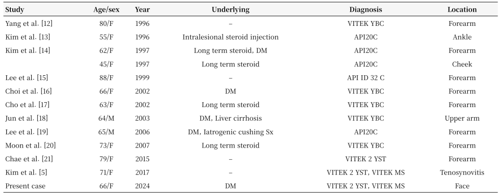
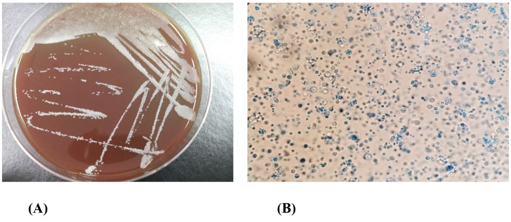
5. Kim JE, Oh TH, Lee KH, Shin JH, Jung SI. Successful treatment of protothecal tenosynovitis in an immunocompetent patient using amphotericin B deoxycholate. Infect Chemother 2017;49:293–6.
6. Todd JR, King JW, Oberle A, Matsumoto T, Odaka Y, Fowler M, et al. Protothecosis: report of a case with 20-year follow-up, and review of previously published cases. Med Mycol 2012;50:673–89.
7. Lass-Flörl C, Mayr A. Human protothecosis. Clin Microbiol Rev 2007;20:230–42.
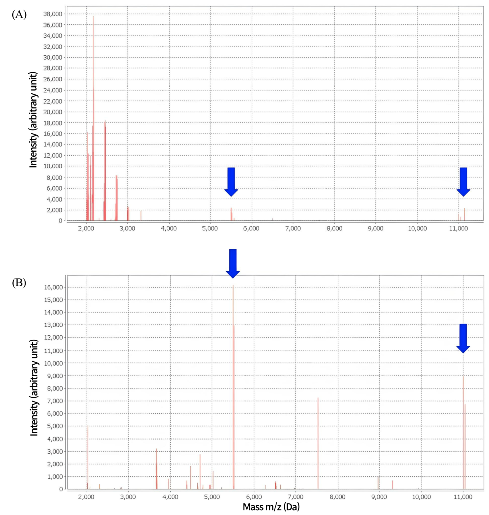
8. Murugaiyan J, Ahrholdt J, Kowbel V, Roesler U. Establishment of a matrix-assisted laser desorption ionization time-of-flight mass spectrometry database for rapid identification of infectious achlorophyllous green micro-algae of the genus Prototheca. Clin Microbiol Infect 2012;18:461–7.
9. Fernández NB, Taverna CG, Vivot M, Córdoba S, Paravano L. First bloodstream infection due to Prototheca zopfii var. hydrocarbonea in an immunocompromised patient. Med Mycol Case Rep 2019;24:9–12.
10. Bizzini A, Jaton K, Romo D, Bille J, Prod’hom G, Greub G. Matrix-assisted laser desorption ionization-time of flight mass spectrometry as an alternative to 16S rRNA gene sequencing for identification of difficult-to-identify bacterial strains. J Clin Microbiol 2011;49:693–6.
11. Sardana R, Butta H, Mendiratta L, Jasuja S, Xess I, Singh G, et al. The balancing universe: the story of human invasion and the primitive yet evolutionary prototheca. Ann Pathol Lab Med 2023;10:132–7.
12. Yang JK, Jang IG, Park YM, Kim TY, Kim HO, Kim CW. A case of cutaneous protothecosis. Ann Dermatol 1996;8:206–10.
13. Kim ST, Suh KS, Chae YS, Kim YJ. Successful treatment with fluconazole of protothecosis developing at the site of an intralesional corticosteroid injection. Br J Dermatol 1996;135:803–6.
14. Kim JA, Moon SE, Song KY. Two cases of cutaneous protothecosis: unique histopathological findings with crystal violet staining and the therapeutic effect of itraconazole. Ann Dermatol 1997;9:201–7.
15. Lee ES, Kim JH, Lee SN. A case of cutaneous protothecosis with severe pustules and ulceration. Kor J Med Mycol 1999;4:131–6.
16. Choi JH, Suh MK, Shin DJ, Suh JC, Yeum JS, Lee HC, et al. A case of cutaneous protothecosis. Korean J Dermatol 2002;40:1116–20.
17. Cho BK, Ham SH, Lee JY, Choi JH. Cutaneous protothecosis. Int J Dermatol 2002;41:304–6.
18. Jun JH, Lee JB, Kim SJ, Lee SC, Won YH. A case of cutaneous protothecosis. Korean J Med Mycol 2003;8:30–4.
19. Lee WS, Kim YJ, Kim SY, Kim KM. A case of cutaneous protothecosis. Korean J Dermatol 2006;44:648–51.
20. Moon HS, Lee HK, Park K, Chae JD, Son SJ. A case of cutaneous protothecosis. Korean J Med Mycol 2007;12:70–4.
21. Chae SY, Lee KC, Lee HS, Jang YH, Lee S, Kim DW, et al. A case of cutaneous protothecosis. Korean J Med Mycol 2015;20:13–8.
22. Croxatto A, Prod’hom G, Greub G. Applications of MALDI-TOF mass spectrometry in clinical diagnostic microbiology. FEMS Microbiol Rev 2012;36:380–407.