Ann Clin Microbiol 2026;29(3):13. https://doi.org/10.5145/ACM.2026.29.3.13
Received on 29 April 2026, Revised on 18 August 2026, Accepted on 26 August 2026, Published on 20 September 2026.
Copyright © Korean Society of Clinical Microbiology.
This is an Open Access article which is freely available under the Creative Commons Attribution-NonCommercial-NoDerivatives 4.0 International License (CC BY-NC-ND) (https://creativecommons.org/licenses/by-nc-nd/4.0/).
Background: Early identification of high-risk patients for severe Clostridioides difficile infection (CDI) is critical for timely intervention. This study evaluated antecedent factors associated with severe CDI while minimizing potential circular reasoning and reverse causality from post-diagnostic clinical course variables.
Methods: In this 10-year retrospective cohort study (2014–2023) at National Health Insurance Service Ilsan Hospital, a Korean referral hospital, 908 consecutive adult inpatients with confirmed toxigenic CDI were enrolled. The patients were categorized as having severe (n = 485) or non-severe (n = 423) CDI according to guideline criteria. Intermediate post-infection events, including intensive care unit admission and total hospital stay, were strictly excluded from predictor models. Univariate screening and multivariable logistic regression were performed using R.
Results: Among 908 patients (median age 78.0 years; 48.6% male), 485 (53.4%) developed severe CDI, which was associated with higher in-hospital mortality than non-severe CDI (26.4% vs. 8.0%, P < 0.001). After multivariable adjustment, independent baseline risk factors for severe CDI were history of prior recurrent CDI (adjusted odds ratio [aOR], 2.77; 95% confidence interval [CI], 1.14–6.77; P = 0.025), prior exposure to β-lactamase inhibitor combinations (aOR, 1.71; 95% CI, 1.25–2.35; P < 0.001), narrow-spectrum cephalosporins (aOR, 2.00; 95% CI, 1.14–3.51; P = 0.016), teicoplanin (aOR, 1.86; 95% CI, 1.08–3.20; P = 0.024), and chronic renal disease (aOR, 1.49; 95% CI, 1.06–2.11; P = 0.022).
Conclusion: A history of recurrent CDI, chronic renal disease, and antecedent exposures to 乒-lactamase inhibitor combinations, narrow-spectrum cephalosporins, and teicoplanin were independently associated with severe CDI, potentially facilitating early risk stratification.
Clostridioides difficile, Risk factors, Severity of illness index, Republic of Korea
Clostridioides difficile is an anaerobic, spore-forming, toxin-producing bacterium that is the primary cause of healthcare-associated infectious colitis worldwide [1]. The clinical spectrum of C. difficile infection (CDI) ranges from self-limiting diarrhea to fulminant pseudomembranous colitis, toxic megacolon, and death [2]. In Korea, widespread use of broad-spectrum antibiotics has increased the incidence and burden of healthcare-associated CDI [3]. Early prediction and risk stratification of severe CDI are essential for timely initiation of targeted oral vancomycin therapy [4]. A recent nationwide survey showed that most Korean physicians tailored CDI treatment according to the disease severity [5].
However, observational studies of CDI frequently suffer from circular reasoning and reverse causality. Variables such as intensive care unit (ICU) admission, length of ICU stay, and prolonged hospitalization are often inappropriately modeled as independent “risk factors” for disease severity. Because ICU admission contributes to the definition of severe-complicated CDI in the severity classification used in this study, ICU-related variables occurring at or after CDI onset cannot serve as temporal antecedent predictors of that outcome and were therefore excluded from the predictor modeling. Moreover, because total hospital stay encompasses the post-diagnostic course, it reflects disease consequences rather than antecedent causes. Eliminating these post-infection variables is important to identify independent baseline factors associated with subsequent severe CDI.
This study aimed to identify antecedent independent risk factors for severe CDI among hospitalized Korean patients over a 10-year period, excluding circular post-infection variables.
This was a single-center, retrospective observational cohort study conducted in accordance with the STROBE guidelines.
This study was conducted at the National Health Insurance Service Ilsan Hospital, an 800-bed public referral hospital in Goyang, Korea. Longitudinal clinical, laboratory, pharmaceutical, and diagnostic data from 2014 to 2023 were extracted from the Ilsan Hospital Data Environment and Analytics (i-DEA) platform.
Eligible participants were consecutive adult inpatients (≥ 19 years) with laboratory-confirmed toxigenic CDI and colitis symptoms. CDI was diagnosed based on laboratory evidence of toxigenic C. difficile using a combination of stool culture, toxin assays, and toxin gene detection using polymerase chain reaction (PCR). C. difficile was cultured on chromID C. difficile agar (bioMérieux), and isolates were identified using matrix-assisted laser desorption/ionization time-of-flight mass spectrometry (Bruker Daltonics). Toxin A/B enzyme immunoassays were performed using either the VIDAS C. difficile Toxin A & B assay (bioMérieux) or the LIAISON C. difficile Toxin A & B assay (DiaSorin Inc.). Real-time PCR targeting tcdB was performed using the Xpert C. difficile assay (Cepheid). To avoid correlated observations, only one index CDI episode per patient was included in the analysis. A history of recurrent CDI was defined as a documented previous CDI episode followed by a new CDI episode occurring 2–8 weeks after the preceding episode and after symptom resolution.
Disease severity was classified according to established criteria [6]. Severe CDI was defined as a serum albumin < 3.0 g/dL plus white blood cell (WBC) count ≥ 15,000/mm³ or abdominal tenderness. Severe-complicated CDI was defined by hypotension, fever ≥ 38.5°C, ileus, mental changes, WBC ≥ 35,000 or < 2,000/mm³, lactate > 2.2 mmol/L, end-organ failure, or ICU admission. Confirmed episodes that met neither definition were categorized as non-severe CDI (controls).
Exposure and confounding variables were distinguished using temporal and causal hierarchies. Exposure variables were defined as antecedent clinical, pharmacological, and environmental factors preceding CDI onset that directly perturb gut microbial ecology or host defenses. These included prior antimicrobial exposures within 12 weeks before diagnosis (narrow-spectrum cephalosporins, extended-spectrum cephalosporins, β-lactamase inhibitor combinations, carbapenems, fluoroquinolones, teicoplanin, penicillin), a history of prior recurrent CDI episodes, healthcare-associated acquisition (symptom onset > 48 h after admission or within 4 weeks of discharge), and concomitant proton pump inhibitor use. Recurrence after the index CDI episode was not included in this variable.
Confounding variables were defined as pre-existing host characteristics and chronic comorbidities that independently influence both the propensity for high-risk antibiotic exposure and host vulnerability to severe infection without acting as direct causal intermediates: baseline age (continuous years), sex (male vs. female), Charlson comorbidity index (CCI) [7], and specific chronic medical conditions verified using standardized diagnostic codes in Korean healthcare databases (chronic renal disease, pneumonia, cerebrovascular disease, malignancy, diabetes mellitus, heart failure, chronic respiratory disease, atherosclerosis, biliary tract disease, liver cirrhosis, and nutritional deficiency) [8]. The CCI was used for descriptive characterization but was not entered simultaneously with individual component comorbidities in the multivariable model to avoid redundant adjustment.
ICU and total hospital stays were classified as post-infection clinical courses and intermediate outcomes, and both were strictly excluded from the multivariable baseline risk-factor association model.
Clinical, pharmacological, and microbiological data were extracted using the i-DEA system. Laboratory values used for severity classification were obtained within ± 3 days of the index CDI test. Because retrospective data availability precludes restricting the analysis to measurements obtained strictly before or during the index test, severity classification may partly reflect early clinical evolution after diagnosis.
The selection bias was minimized by enrolling consecutive inpatients. Information bias was mitigated using electronic records. The potential for circular reasoning and reverse causality was minimized by restricting the predictors to antecedent events and excluding post-diagnostic ICU- and hospital-stay variables.
All eligible patients enrolled consecutively over a 10-year period (N = 908) were included. With 485 severe CDI events, the dataset provided sufficient outcome events relative to the number of parameters included in the final multivariate model.
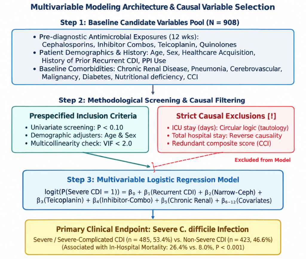
Continuous variables are presented as median (interquartile range) or mean (standard deviation) and compared using the Mann–Whitney U test or Welch t-test. Categorical variables are expressed as frequencies and percentages and were compared using the chi-square or Fisher’s exact test. The unadjusted odds ratios (ORs) and 95% confidence intervals (CIs) were calculated. Candidate baseline predictor variables with P < 0.10 in univariate analysis, along with age and sex, were entered into multivariable logistic regression. Multicollinearity was evaluated using variance inflation factors (all < 2.0). The tests were two-tailed (P < 0.05). The overall multivariable modeling architecture, prespecified screening criteria, and strict causal exclusion of post-infection circular variables are shown in Fig. 1. Analyses were performed using the R software version 4.4.2 (R Foundation for Statistical Computing).
Fig. 1. Multivariable modeling architecture and causal variable selection framework. Overview of candidate predictor pool (n = 908), prespecified inclusion criteria (univariate P < 0.10, age and sex adjustment, VIF < 2.0), strict causal exclusions (eliminating circular ICU stay, total hospital stay, and collinear composite CCI scores), and final logistic model formulation targeting severe Clostridioides difficile infection (CDI).
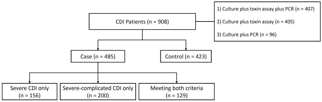
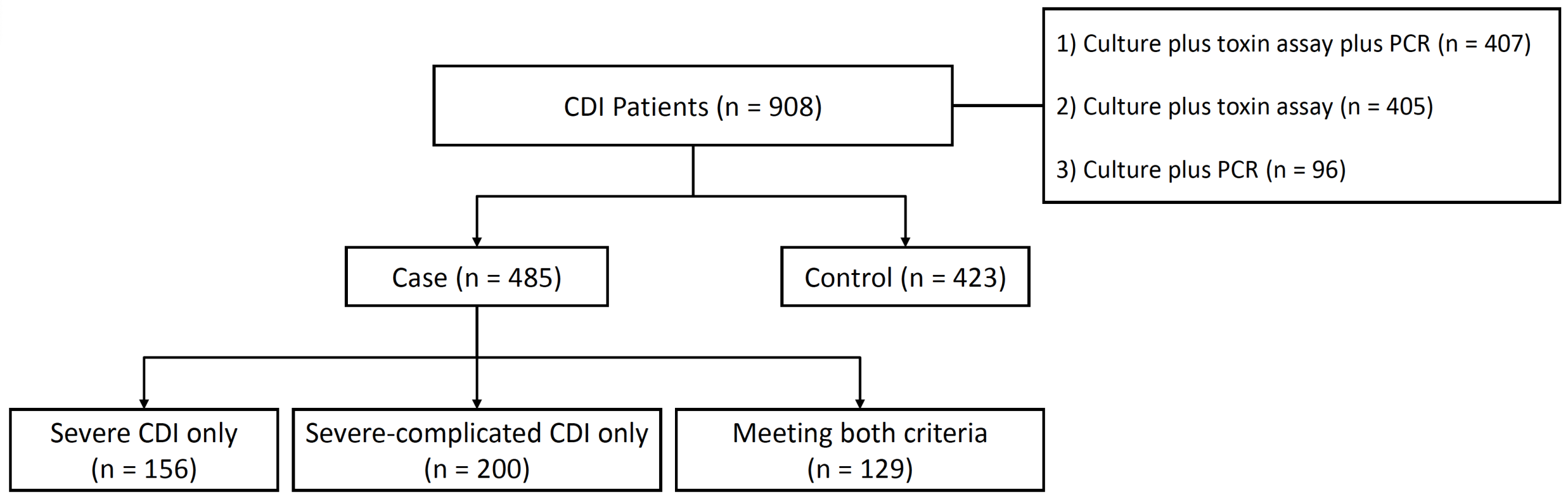
Over 10 years, 908 unique adult inpatients with confirmed CDI were enrolled (Fig. 2). Non-severe CDI accounted for 423 patients (46.6%) and severe CDI for 485 patients (53.4%). Among the severe cases, 156 met the criteria for severe CDI only, 200 met the criteria for severe-complicated CDI only, and 129 met both criteria, totaling 329 patients (36.2%) with severe-complicated features.
Fig. 2. Flowchart of patient enrollment and disease severity stratification. Patients with confirmed CDI (n = 908) were stratified into non-severe CDI controls (n = 423) and severe CDI cases (n = 485). Severe cases comprised three mutually exclusive subgroups: severe CDI only (n = 156), severe-complicated CDI only (n = 200), and patients meeting both sets of criteria (n = 129). CDI, Clostridioides difficile infection; PCR, polymerase chain reaction.
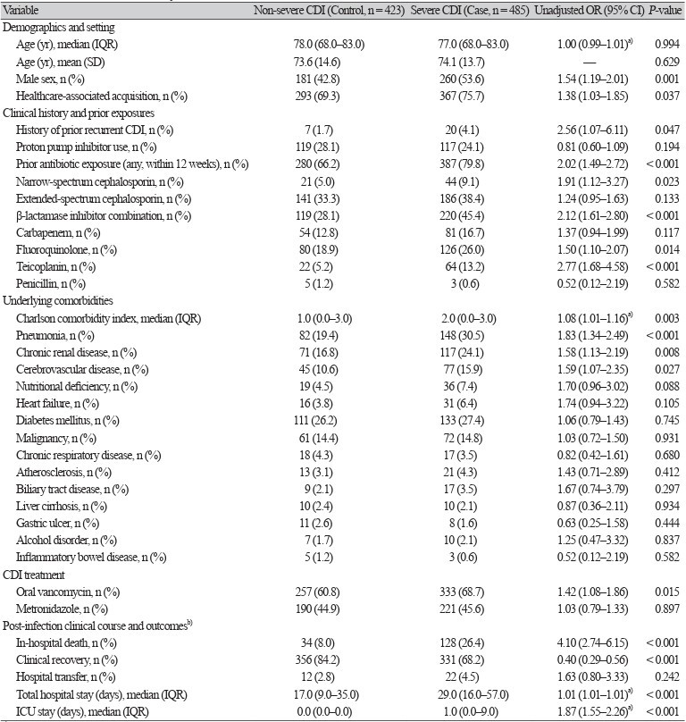
The baseline characteristics and unadjusted comparisons between non-severe controls and severe cases are presented in Table 1. Severe CDI cases had significantly higher proportions of male sex, healthcare-associated acquisition, and prior recurrent CDI history. Pre-diagnostic exposure to antimicrobials was significantly more prevalent among severe cases, driven by higher utilization of narrow-spectrum cephalosporins, β-lactamase inhibitor combinations, fluoroquinolones, and teicoplanin. Pneumonia, chronic renal disease, cerebrovascular disease, and higher overall comorbidity scores were significantly more frequent in the severe cases. Patients with severe CDI also had markedly worse clinical outcomes, including higher in-hospital mortality (26.4% vs. 8.0%, P < 0.001), lower clinical recovery, and significantly prolonged hospital stays.
Table 1. Baseline demographic, clinical, and pharmacological characteristics of patients with non-severe and severe Clostridioides difficile infection: univariate analysis
| Variable | Non-severe CDI (Control, n = 423) | Severe CDI (Case, n = 485) | Unadjusted OR (95% CI) | P-value |
|---|---|---|---|---|
| Demographics and setting | ||||
| Age (yr), median (IQR) | 78.0 (68.0–83.0) | 77.0 (68.0–83.0) | 1.00 (0.99–1.01)a) | 0.994 |
| Age (yr), mean (SD) | 73.6 (14.6) | 74.1 (13.7) | — | 0.629 |
| Male sex, n (%) | 181 (42.8) | 260 (53.6) | 1.54 (1.19–2.01) | 0.001 |
| Healthcare-associated acquisition, n (%) | 293 (69.3) | 367 (75.7) | 1.38 (1.03–1.85) | 0.037 |
| Clinical history and prior exposures | ||||
| History of prior recurrent CDI, n (%) | 7 (1.7) | 20 (4.1) | 2.56 (1.07–6.11) | 0.047 |
| Proton pump inhibitor use, n (%) | 119 (28.1) | 117 (24.1) | 0.81 (0.60–1.09) | 0.194 |
| Prior antibiotic exposure (any, within 12 weeks), n (%) | 280 (66.2) | 387 (79.8) | 2.02 (1.49–2.72) | < 0.001 |
| Narrow-spectrum cephalosporin, n (%) | 21 (5.0) | 44 (9.1) | 1.91 (1.12–3.27) | 0.023 |
| Extended-spectrum cephalosporin, n (%) | 141 (33.3) | 186 (38.4) | 1.24 (0.95–1.63) | 0.133 |
| β-lactamase inhibitor combination, n (%) | 119 (28.1) | 220 (45.4) | 2.12 (1.61–2.80) | < 0.001 |
| Carbapenem, n (%) | 54 (12.8) | 81 (16.7) | 1.37 (0.94–1.99) | 0.117 |
| Fluoroquinolone, n (%) | 80 (18.9) | 126 (26.0) | 1.50 (1.10–2.07) | 0.014 |
| Teicoplanin, n (%) | 22 (5.2) | 64 (13.2) | 2.77 (1.68–4.58) | < 0.001 |
| Penicillin, n (%) | 5 (1.2) | 3 (0.6) | 0.52 (0.12–2.19) | 0.582 |
| Underlying comorbidities | ||||
| Charlson comorbidity index, median (IQR) | 1.0 (0.0–3.0) | 2.0 (0.0–3.0) | 1.08 (1.01–1.16)a) | 0.003 |
| Pneumonia, n (%) | 82 (19.4) | 148 (30.5) | 1.83 (1.34–2.49) | < 0.001 |
| Chronic renal disease, n (%) | 71 (16.8) | 117 (24.1) | 1.58 (1.13–2.19) | 0.008 |
| Cerebrovascular disease, n (%) | 45 (10.6) | 77 (15.9) | 1.59 (1.07–2.35) | 0.027 |
| Nutritional deficiency, n (%) | 19 (4.5) | 36 (7.4) | 1.70 (0.96–3.02) | 0.088 |
| Heart failure, n (%) | 16 (3.8) | 31 (6.4) | 1.74 (0.94–3.22) | 0.105 |
| Diabetes mellitus, n (%) | 111 (26.2) | 133 (27.4) | 1.06 (0.79–1.43) | 0.745 |
| Malignancy, n (%) | 61 (14.4) | 72 (14.8) | 1.03 (0.72–1.50) | 0.931 |
| Chronic respiratory disease, n (%) | 18 (4.3) | 17 (3.5) | 0.82 (0.42–1.61) | 0.680 |
| Atherosclerosis, n (%) | 13 (3.1) | 21 (4.3) | 1.43 (0.71–2.89) | 0.412 |
| Biliary tract disease, n (%) | 9 (2.1) | 17 (3.5) | 1.67 (0.74–3.79) | 0.297 |
| Liver cirrhosis, n (%) | 10 (2.4) | 10 (2.1) | 0.87 (0.36–2.11) | 0.934 |
| Gastric ulcer, n (%) | 11 (2.6) | 8 (1.6) | 0.63 (0.25–1.58) | 0.444 |
| Alcohol disorder, n (%) | 7 (1.7) | 10 (2.1) | 1.25 (0.47–3.32) | 0.837 |
| Inflammatory bowel disease, n (%) | 5 (1.2) | 3 (0.6) | 0.52 (0.12–2.19) | 0.582 |
| CDI treatment | ||||
| Oral vancomycin, n (%) | 257 (60.8) | 333 (68.7) | 1.42 (1.08–1.86) | 0.015 |
| Metronidazole, n (%) | 190 (44.9) | 221 (45.6) | 1.03 (0.79–1.33) | 0.897 |
| Post-infection clinical course and outcomesb) | ||||
| In-hospital death, n (%) | 34 (8.0) | 128 (26.4) | 4.10 (2.74–6.15) | < 0.001 |
| Clinical recovery, n (%) | 356 (84.2) | 331 (68.2) | 0.40 (0.29–0.56) | < 0.001 |
| Hospital transfer, n (%) | 12 (2.8) | 22 (4.5) | 1.63 (0.80–3.33) | 0.242 |
| Total hospital stay (days), median (IQR) | 17.0 (9.0–35.0) | 29.0 (16.0–57.0) | 1.01 (1.01–1.01)a) | < 0.001 |
| ICU stay (days), median (IQR) | 0.0 (0.0–0.0) | 1.0 (0.0–9.0) | 1.87 (1.55–2.26)a) | < 0.001 |
a)Odds ratios for continuous variables (age, Charlson comorbidity index, hospital stay, ICU stay) are presented per 1-unit increase.
b)Intermediate clinical course and post-infection outcomes (hospital stay, ICU stay, in-hospital death) are presented for descriptive characterization but were strictly excluded from multivariable prediction models to prevent circular logic and reverse causality.
Abbreviations: CDI, Clostridioides difficile infection; OR, odds ratio; CI, confidence interval; IQR, interquartile range; SD, standard deviation; ICU, intensive care unit.
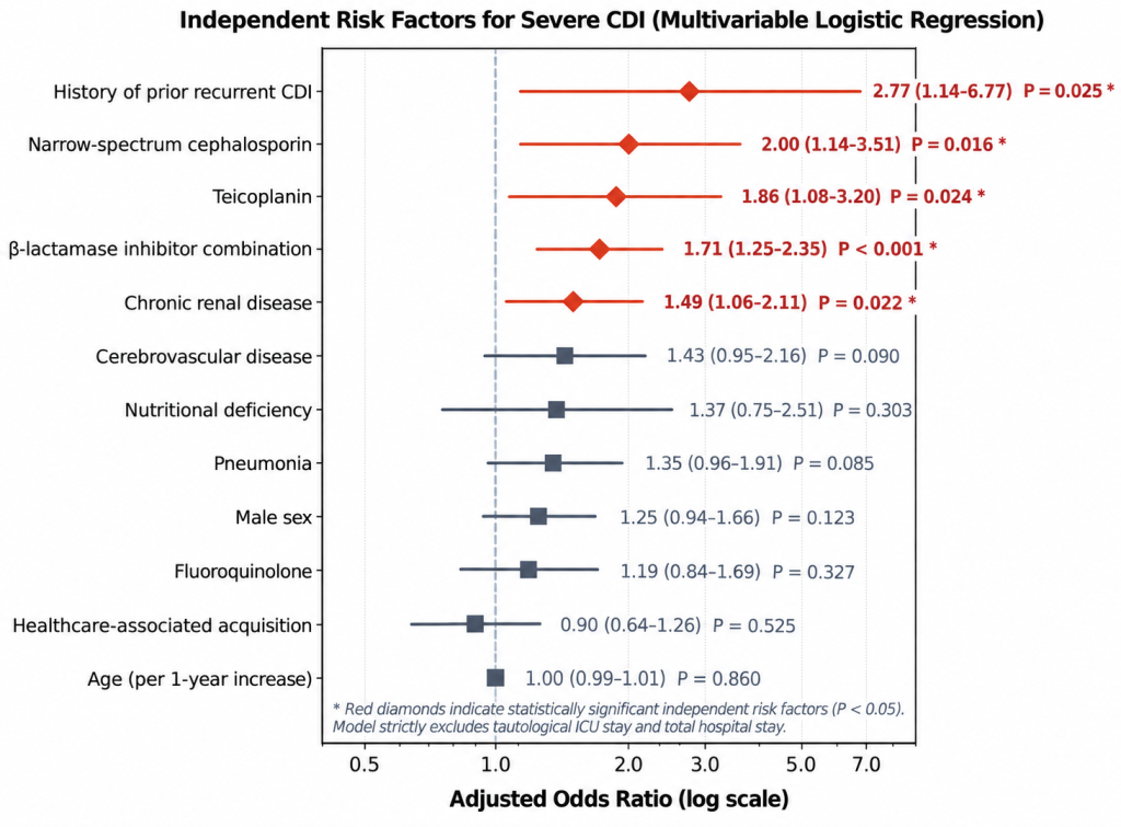
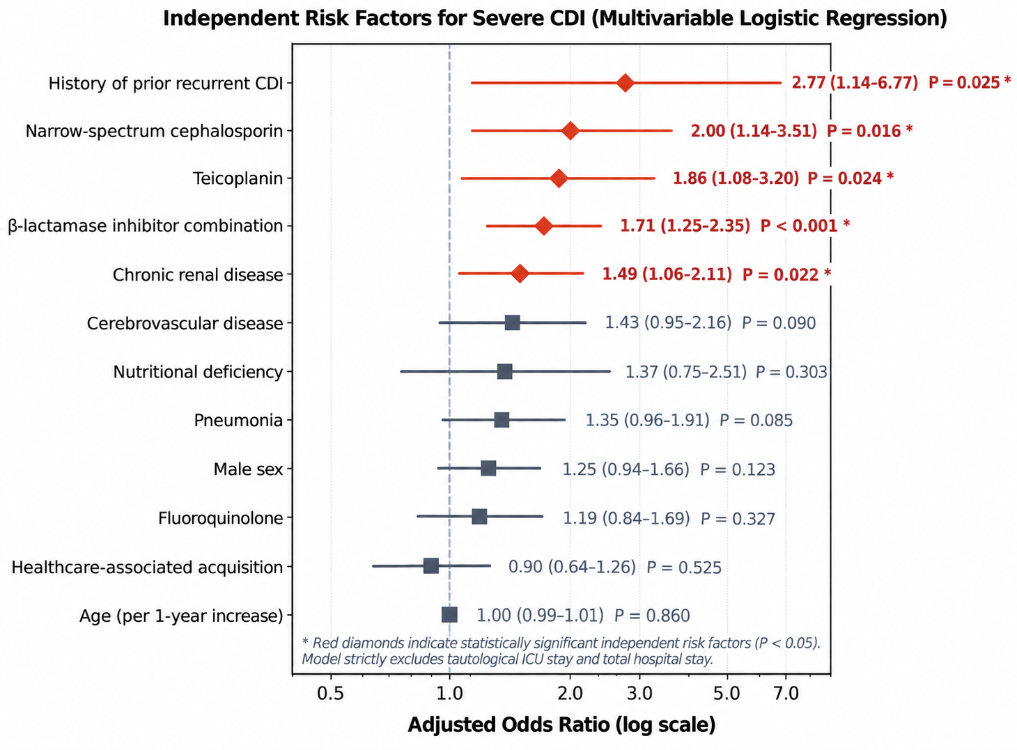
In multivariable logistic regression modeling (Fig. 3), five factors emerged as statistically significant independent baseline predictors of severe CDI: history of prior recurrent CDI (adjusted OR [aOR], 2.77; 95% CI, 1.14–6.77; P = 0.025), prior exposure to narrow-spectrum cephalosporins (aOR, 2.00; 95% CI, 1.14–3.51; P = 0.016), teicoplanin (aOR, 1.86; 95% CI, 1.08–3.20; P = 0.024), β-lactamase inhibitor combinations (aOR, 1.71; 95% CI, 1.25–2.35; P < 0.001), and chronic renal disease (aOR, 1.49; 95% CI, 1.06–2.11; P = 0.022). Borderline associations were observed for cerebrovascular disease (aOR, 1.43; 95% CI, 0.95–2.16; P = 0.090) and pneumonia (aOR, 1.35; 95% CI, 0.96–1.91; P = 0.085).
Fig. 3. Independent risk factors for severe Clostridioides difficile infection. Forest plot of adjusted odds ratios (aOR) and 95% confidence intervals (CI) from multivariable logistic regression analysis. Bold red diamonds indicate statistically significant independent baseline risk factors (P < 0.05).
In this 10-year cohort of 908 Korean inpatients with toxigenic CDI, severe disease was associated with substantially higher in-hospital mortality (26.4% vs. 8.0%; unadjusted OR, 4.10). After excluding post-diagnostic hospital stay and ICU duration variables from the model, multivariable modeling demonstrated that prior history of recurrent CDI, pre-diagnostic exposure to narrow-spectrum cephalosporins, β-lactamase inhibitor combinations, or teicoplanin, and underlying chronic renal disease were independent baseline risk factors for severe CDI.
These findings provide essential epidemiological clarity by correcting the circular logic presented in the earlier literature. When ICU stay is included as an explanatory variable, it artificially dominates regression models because ICU transfer forms part of the severe-complicated disease definition. By appropriately classifying post-infection hospital metrics as outcome measures, this approach reduces the potential for reverse causality arising from the inclusion of post-diagnostic clinical course variables.
A history of recurrent CDI may reflect persistent microbiome disruption and host susceptibility. However, the biological mechanisms underlying its association with subsequent severe presentation could not be determined in this retrospective study. The association with teicoplanin may reflect both microbiome disruption and confounding by indication, because teicoplanin is preferentially administered to patients with serious Gram-positive infections and greater baseline clinical complexity. Furthermore, chronic renal disease may contribute to systemic immune dysfunction, uremic enteropathy, and delayed toxin clearance, potentially accelerating disease progression.
This study offers several distinct methodological and epidemiological strengths compared with previously published CDI studies. First, our analysis provided full transparency by reporting both comprehensive univariate screening and mutually adjusted multivariate logistic regression across all clinical variables, resolving the reporting opacity common in retrospective studies. Second, rather than crudely grouping all antimicrobial agents into an undifferentiated broad-spectrum category, our study demonstrates class-specific vulnerability, uncovering substantial independent risks specifically for β-lactamase inhibitor combinations, narrow-spectrum cephalosporins, and teicoplanin.
In this study, the in-hospital mortality in patients with severe CDI was 26.4%. A recent Korean multicenter study also documented a substantial burden of adverse outcomes in severe CDI, with approximately one-quarter of the patients experiencing all-cause death or colectomy within 60 days of diagnosis [9]. Furthermore, severe systemic inflammatory responses and poor patient outcomes have been linked to severe clostridial illness in Korean acute-care settings [10]. Our findings also reflect the substantial inpatient healthcare burden and prolonged hospitalization documented in nationwide economic evaluations [11].
This study has several limitations. First, it was conducted at a single secondary referral hospital, which may limit its generalizability to community settings. Second, molecular ribotyping and binary toxin analysis have not been routinely performed across all years, although recent surveillance has indicated substantial genetic diversity among Korean clinical isolates [12]. Third, the precise cumulative antibiotic doses could not be quantified. Nevertheless, the consecutive enrollment of 908 microbiologically confirmed cases over a decade and the separation of antecedent exposures from post-diagnostic clinical course variables strengthened the internal validity of the analysis. Fourth, the ±3-day window used to retrieve laboratory parameters may have captured early post-diagnostic changes in disease severity. Therefore, although the predictor variables were restricted to antecedent exposures, the outcome should not be interpreted strictly as severity at the time of CDI diagnosis. Finally, residual confounding by indication is possible because antecedent antimicrobial exposure may partly reflect the severity and complexity of the underlying infections.
The study population, which included a large proportion of older and medically complex inpatients, may be broadly representative of the patients encountered in Korean acute-care hospitals. Nevertheless, the single-center design warrants caution when extrapolating these findings to other healthcare settings.
Clinicians should maintain heightened vigilance regarding severe CDI when treating patients with chronic kidney disease, prior CDI history, or recent exposure to β-lactamase inhibitor combinations, narrow-spectrum cephalosporins, or teicoplanin. These high-risk individuals may warrant closer clinical monitoring and prompt assessment of CDI severity to guide treatment in accordance with the current clinical guidelines. These findings support continued antimicrobial stewardship and appropriate de-escalation of broad-spectrum antimicrobial therapy when clinically feasible.
Severe CDI is associated with substantial in-hospital mortality in hospitalized Korean patients. After excluding post-diagnostic hospital-course variables from the multivariable model, antecedent history of recurrent CDI, chronic renal disease, and prior exposures to β-lactamase inhibitor combinations, narrow-spectrum cephalosporins, and teicoplanin were independently associated with severe CDI after adjustment for measured baseline covariates. These pre-diagnostic factors may help identify patients who warrant closer assessment for severe CDI, although their utility for prospective risk stratification requires further validation.
The Institutional Review Board of the National Health Insurance Service Ilsan Hospital approved this retrospective cohort study (approval number: NHIMC 2024-03-043) and waived the requirement for informed consent because anonymized data were used.
No potential conflict of interest relevant to this article was reported.
This work was supported by the National Health Insurance Service Ilsan Hospital research fund (grant number: NHIMC-2024-CR-051). The funders had no role in study design, data collection and analysis, decision to publish, or preparation of the manuscript.
The datasets generated during the current study are available from the corresponding author upon request.
We thank Hayeon Kim (Department of Medical Records, National Health Insurance Service Ilsan Hospital) for assistance with clinical data extraction, and Hyunsun Lim (Department of Research and Analysis, National Health Insurance Service Ilsan Hospital) for advice on data curation.
1. Salvati F, Catania F, Murri R, Fantoni M, Torti C. Clostridioides difficile infection: an update. Infez Med 2024;32:280-91.

2. Jeon SR. The burden of Clostridioides difficile infection in Korea. J Korean Med Sci 2024;39:e122.

3. Kim J, Myung R, Kim B, Kim J, Kim T, Lee MS, et al. Incidence of Clostridioides difficile infections in Republic of Korea: a prospective study with active surveillance vs. national data from Health Insurance Review & Assessment Service. J Korean Med Sci 2024;39:e118.

4. Cymbal M, Chatterjee A, Baggott B, Auron M. Management of Clostridioides difficile infection: diagnosis, treatment, and future perspectives. Am J Med 2024;137:571-6.

5. Moon JM, Kim SE, Kim J, Cho YS, Kim H, Gweon TG, et al. Clinical approaches to Clostridioides difficile infection management: insights from a nationwide survey of Korean physicians. J Korean Med Sci 2026;41:e150.

6. Surawicz CM, Brandt LJ, Binion DG, Ananthakrishnan AN, Curry SR, Gilligan PH, et al. Guidelines for the diagnosis, treatment, and prevention of Clostridium difficile infections. Am J Gastroenterol 2013;108:478-98.

7. Soos B, Williamson T, McBrien K, Wiebe S, Tonelli M, Southern DA, et al. Considerations for selecting and implementing comorbidity indices when using secondary data sources: a guide for health researchers. Int J Popul Data Sci 2025;10:2973.

8. Korean Institute of Medical Classification and Information Center (KOICD). Integrated search for disease classification codes. https://www.koicd.kr/sch/searchTotal.do [Online] (last visited on 13 September 2026).
9. Cho YW, Moon JM, Lee HH, Kim J, Choi CH, Lee KM, et al. Combination therapy with oral vancomycin plus intravenous metronidazole is not superior to oral vancomycin alone for the treatment of severe Clostridioides difficile infection: a KASID multicenter study. Antibiotics (Basel) 2025;14:1252.

10. Choi IH, Kim I, Lee HH, Kim J, Cho YS. Eosinopenia is associated with worse outcomes in hospitalized patients with Clostridioides difficile infection in South Korea. Anaerobe 2026;98:103047.

11. Myung R, Lee E, Kim J, Kim J, Pai H. Attributable costs of Clostridioides difficile infections in Korea. J Korean Med Sci 2025;40:e22.

12. Kim J, Kim B, Pai H. Diversity of binary toxin positive Clostridioides difficile in Korea. Sci Rep 2023;13:576.
1. Salvati F, Catania F, Murri R, Fantoni M, Torti C. Clostridioides difficile infection: an update. Infez Med 2024;32:280-91.

2. Jeon SR. The burden of Clostridioides difficile infection in Korea. J Korean Med Sci 2024;39:e122.

3. Kim J, Myung R, Kim B, Kim J, Kim T, Lee MS, et al. Incidence of Clostridioides difficile infections in Republic of Korea: a prospective study with active surveillance vs. national data from Health Insurance Review & Assessment Service. J Korean Med Sci 2024;39:e118.

4. Cymbal M, Chatterjee A, Baggott B, Auron M. Management of Clostridioides difficile infection: diagnosis, treatment, and future perspectives. Am J Med 2024;137:571-6.

5. Moon JM, Kim SE, Kim J, Cho YS, Kim H, Gweon TG, et al. Clinical approaches to Clostridioides difficile infection management: insights from a nationwide survey of Korean physicians. J Korean Med Sci 2026;41:e150.

6. Surawicz CM, Brandt LJ, Binion DG, Ananthakrishnan AN, Curry SR, Gilligan PH, et al. Guidelines for the diagnosis, treatment, and prevention of Clostridium difficile infections. Am J Gastroenterol 2013;108:478-98.

7. Soos B, Williamson T, McBrien K, Wiebe S, Tonelli M, Southern DA, et al. Considerations for selecting and implementing comorbidity indices when using secondary data sources: a guide for health researchers. Int J Popul Data Sci 2025;10:2973.

8. Korean Institute of Medical Classification and Information Center (KOICD). Integrated search for disease classification codes. https://www.koicd.kr/sch/searchTotal.do [Online] (last visited on 13 September 2026).
9. Cho YW, Moon JM, Lee HH, Kim J, Choi CH, Lee KM, et al. Combination therapy with oral vancomycin plus intravenous metronidazole is not superior to oral vancomycin alone for the treatment of severe Clostridioides difficile infection: a KASID multicenter study. Antibiotics (Basel) 2025;14:1252.

10. Choi IH, Kim I, Lee HH, Kim J, Cho YS. Eosinopenia is associated with worse outcomes in hospitalized patients with Clostridioides difficile infection in South Korea. Anaerobe 2026;98:103047.

11. Myung R, Lee E, Kim J, Kim J, Pai H. Attributable costs of Clostridioides difficile infections in Korea. J Korean Med Sci 2025;40:e22.

12. Kim J, Kim B, Pai H. Diversity of binary toxin positive Clostridioides difficile in Korea. Sci Rep 2023;13:576.