Table 1. Baseline demographic, clinical, and pharmacological characteristics of patients with non-severe and severe Clostridioides difficile infection: univariate analysis
Ann Clin Microbiol 2026;29(3):13. Risk factors for severe Clostridioides difficile infection in Korea: a retrospective cohort study Download table Variable Non-severe CDI (Control, n = 423) Severe CDI (Case, n = 485) Unadjusted OR (95% CI) P-value Demographics and setting Age (yr), median (IQR) 78.0 (68.0–83.0) 77.0 (68.0–83.0) 1.00 (0.99–1.01)a) 0.994 Age […]
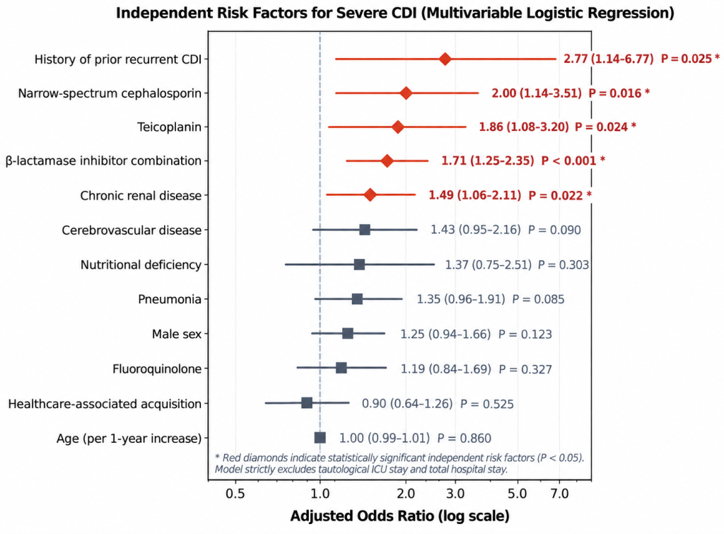
Fig. 3. Independent risk factors for severe Clostridioides difficile infection. Forest plot of adjusted odds ratios (aOR) and 95% confidence intervals (CI) from multivariable logistic regression analysis. Bold red diamonds indicate statistically significant independent baseline risk factors (P < 0.05).
Ann Clin Microbiol 2026;29(3):13. Risk factors for severe Clostridioides difficile infection in Korea: a retrospective cohort study Download image
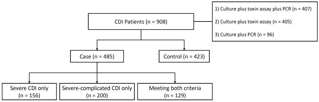
Fig. 2. Flowchart of patient enrollment and disease severity stratification. Patients with confirmed CDI (n = 908) were stratified into non-severe CDI controls (n = 423) and severe CDI cases (n = 485). Severe cases comprised three mutually exclusive subgroups: severe CDI only (n = 156), severe-complicated CDI only (n = 200), and patients meeting both sets of criteria (n = 129). CDI, Clostridioides difficile infection; PCR, polymerase chain reaction.
Ann Clin Microbiol 2026;29(3):13. Risk factors for severe Clostridioides difficile infection in Korea: a retrospective cohort study Download image
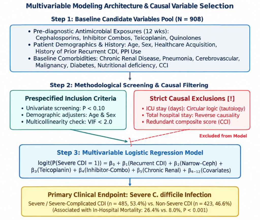
Fig. 1. Multivariable modeling architecture and causal variable selection framework. Overview of candidate predictor pool (n = 908), prespecified inclusion criteria (univariate P < 0.10, age and sex adjustment, VIF < 2.0), strict causal exclusions (eliminating circular ICU stay, total hospital stay, and collinear composite CCI scores), and final logistic model formulation targeting severe Clostridioides difficile infection (CDI).
Ann Clin Microbiol 2026;29(3):13. Risk factors for severe Clostridioides difficile infection in Korea: a retrospective cohort study Download image
Risk factors for severe Clostridioides difficile infection in Korea: a retrospective cohort study
Review article Banseok Kim , Young Ah Kim Department of Laboratory Medicine, National Health Insurance Service Ilsan Hospital, Goyang, Korea Ann Clin Microbiol 2026;29(3):13. https://doi.org/10.5145/ACM.2026.29.3.13Received on 29 April 2026, Revised on 18 August 2026, Accepted on 26 August 2026, Published on 20 September 2026.Copyright © Korean Society of Clinical Microbiology.This is an Open Access article […]
Table 3. Antimicrobial resistance and gene patterns of Salmonella Typhimurium variants
Ann Clin Microbiol 2026;29(3):14. Molecular epidemiological characteristics of monophasic Salmonella enterica serovar Typhimurium variants in Korea Download table Antimicrobial agent I 4,[5],12:i:1,2 (Typhimurium) I 4,[5],12:-:- I 4,[5],12:i:- P1(n = 32) P2(n = 6) Total(n = 38) P1(n = 4) P1(n = 2) P5(n = 72) P8(n = 29) P9(n = 28) Total(n […]
Table 2. Antigenic formula and gene pattern of Salmonella Typhimurium variants
Ann Clin Microbiol 2026;29(3):14. Molecular epidemiological characteristics of monophasic Salmonella enterica serovar Typhimurium variants in Korea Download table Gene patterna) Antigenic formula (n, %) Total I 4,[5],12:-:- I 4,[5],12:i:- I 4,[5],12:i:1,2 (Typhimurium) P1 (+/+/+/+/+/+/+) 4 (100.0) 2 (1.5) 32 (84.2) 38 (22.0) P2 (+/+/−/+/+/+/+) − − 6 (15.8) 6 (3.5) P5 (+/+/+/+/−/−/−) − 72 (55.0) − […]
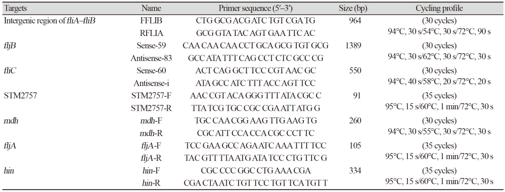
Table 1. Primers and polymerase chain reaction conditions used in this study
Ann Clin Microbiol 2026;29(3):14. Molecular epidemiological characteristics of monophasic Salmonella enterica serovar Typhimurium variants in Korea Download table Targets Name Primer sequence (5′–3′) Size (bp) Cycling profile Intergenic region of fliA–fliB FFLIB CTG GCG ACG ATC TGT CGA TG 964 (30 cycles)94℃, 30 s/54℃, 30 s/72℃, 90 s RFLIA GCG GTA TAC AGT GAA TTC AC […]
Molecular epidemiological characteristics of monophasic Salmonella enterica serovar Typhimurium variants in Korea
Original article Eun-Young Kim1,2 , Su Man Kim1,2 , Si Hyun Kim3 , Jeong Hwan Shin1,2 1Department of Laboratory Medicine, Inje University Busan Paik Hospital, Inje University College of Medicine, Busan, Korea2Paik Institute for Clinical Research, Inje University College of Medicine, Busan, Korea3Department of Biomedical Laboratory Science, Inje University, Gimhae, Korea Ann Clin Microbiol 2026;29(3):14. […]
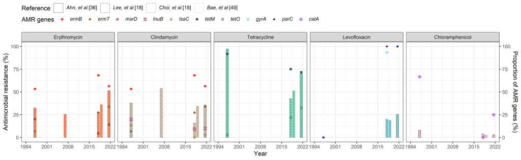
Fig. 1. Antimicrobial resistance (AMR) profiles and associated genes of group B Streptococcus (GBS) isolated from South Korean women of childbearing age and pregnant women. The x-axis shows the reference studies included in the analysis and the corresponding bacterial isolate collection periods. Each column uses a distinct border to distinguish individual studies and is positioned at the midpoint of the isolate collection period for that study. Column heights represent phenotypic antimicrobial resistance rates (left y-axis). Distinct geometric symbols represent the proportions of isolates carrying AMR genes or resistance-associated mutations among GBS isolates resistant to the corresponding antimicrobial agent in each study (right y-axis). Estimates were derived from independent studies with heterogeneous populations, sampling strategies, and laboratory methods and therefore should not be interpreted as a continuous longitudinal national trend.
Ann Clin Microbiol 2026;29(3):12. Evolving molecular epidemiology and antimicrobial resistance of Streptococcus agalactiae in South Korea and East Asia: a narrative review Download image