Annals of Clinical Microbiology, The official Journal of the Korean Society of Clinical Microbiology

6

Weeks in Review

4

Weeks to Publication
Indexed in KCI, KoreaMed, Synapse, DOAJ
Open Access, Peer Reviewed
pISSN 2288-0585 eISSN 2288-6850
Original article

Molecular epidemiological characteristics of monophasic Salmonella enterica serovar Typhimurium variants in Korea

1Department of Laboratory Medicine, Inje University Busan Paik Hospital, Inje University College of Medicine, Busan, Korea
2Paik Institute for Clinical Research, Inje University College of Medicine, Busan, Korea
3Department of Biomedical Laboratory Science, Inje University, Gimhae, Korea

Ann Clin Microbiol 2026;29(3):14. https://doi.org/10.5145/ACM.2026.29.3.14
Received on 21 July 2026, Revised on 1 September 2026, Accepted on 10 September 2026, Published on 20 September 2026.
Copyright © Korean Society of Clinical Microbiology.
This is an Open Access article which is freely available under the Creative Commons Attribution-NonCommercial-NoDerivatives 4.0 International License (CC BY-NC-ND) (https://creativecommons.org/licenses/by-nc-nd/4.0/).

Abstract

Background: Monophasic Salmonella enterica serovar Typhimurium variants, particularly the serotype I 4,[5],12:i:-, have globally emerged as important foodborne pathogens associated with multidrug resistance. However, their molecular epidemiology and clonal distribution in South Korea remain unclear. This study investigated the distribution, clonal characteristics, and antimicrobial resistance profiles of S. Typhimurium variants circulating in South Korea.

Methods: In total, 173 S. Typhimurium variant strains were collected from 18 university hospitals between 2019 and 2020. Isolates were identified and serotyped according to the Kauffmann〝White scheme. Gene patterns were determined based on seven phase 1 and 2 flagellar antigen-associated genes. Antimicrobial susceptibility was examined using broth microdilution.

Results: The monophasic variant I 4,[5],12:i:- (n = 131) was the most prevalent, followed by I 4,[5],12:i:1,2 (n = 38) and I 4,[5],12:-:- (n = 4). Gene patterns dominant among monophasic isolates included P5 (n = 72), P8 (n = 29), and P9 (n = 28). The P5 pattern corresponded to the typical Spanish clone. Monophasic variants showed higher resistance rates to ampicillin and third-generation cephalosporins than those of other variants, with higher cephalosporin resistance observed in P8 than in P5.

Conclusion: Among the isolates included in this study, the monophasic variant I 4,[5],12:i:- was more frequently identified than typical S. Typhimurium. Most I 4,[5],12:i:- isolates showed gene patterns previously associated with the Spanish clone and high rates of antimicrobial resistance. Overall, these findings highlight the need for continued surveillance of I 4,[5],12:i:- and its antimicrobial resistance patterns.

Keywords

Drug Resistance, Bacterial; Molecular epidemiology; Salmonella Typhimurium

Introduction

Background

Salmonella enterica is one of the leading causes of foodborne bacterial illness worldwide [1], with > 2,500 serotypes identified using the Kauffmann–White scheme [2]. The epidemiology and clinical severity of salmonellosis vary with serotype, and certain serotypes are associated with specific reservoirs and severe disease in humans [3,4].

Salmonella enterica serovar Typhimurium (antigenic formula: I 4,[5],12:i:1,2) is one of the most prevalent serotypes worldwide. However, human infections caused by its variants, including monophasic variant (I 4,[5],12:i:-), have increased [57]. Accurately identifying these variants is important as they may differ in antimicrobial resistance and transmission patterns.

To improve the identification of S. Typhimurium variants, the European Food Safety Authority (EFSA) has proposed the molecular approaches targeting the phase 1 and phase 2 flagellar genes by Tennant et al. [8]. Further, Bugarel et al. [9] have proposed a refined polymerase chain reaction (PCR)-based classification system that defined 11 gene patterns (P1–P11).

S. Typhimurium variants, including I 4,[5],12:i:-, are increasing in Asia including Japan and South Korea [57]; however, the data on their molecular epidemiology and clonal distribution are limited.

Objectives

This study aimed to investigate the distribution and molecular epidemiological characteristics of S. enterica serovar Typhimurium variants in South Korea.

Methods

Study design

This retrospective surveillance study was based on laboratory investigations. The study was undertaken according to the Microbiology Investigation Criteria for Reporting Objectively (MICRO) framework for the reporting and interpretation of clinical microbiology data [10].

Laboratory studies

Bacterial isolates

In total, this study included 173 S. Typhimurium variant strains collected from 18 hospitals across South Korea between 2019 and 2020. The participating hospitals were located in Seoul, Incheon, Gyeonggi, Gangwon, Chungcheong, Daegu, Ulsan, Busan, Gwangju, Jeolla, and Jeju. All isolates were recovered from clinical specimens submitted for diagnostic testing and were not obtained through non-diagnostic surveillance or screening. The specimen sources included stool (n = 126), blood (n = 36), and other specimens (n = 11).

At each participating hospital, isolates recovered from clinical specimens were stored in 10% skim milk at −70°C and transported frozen to Inje University Busan Paik Hospital, where all subsequent experiments were performed.

The analysis included only the first isolate of each Salmonella enterica serovar Typhimurium variant obtained from each patient during the study period. Duplicate isolates from the same patient were excluded.

Identification and serotyping

All isolates were confirmed as S. enterica using the VITEK MS system with database version 3.0 (bioMérieux SA). Serotyping was performed using slide agglutination with somatic (O) and flagellar (H) antisera (BD Difco™, Becton, Dickinson and Company), and antigenic formulas were determined according to the Kauffmann–White scheme [11]. Phase inversion was performed to assess phase 2 H-antigen expression and to distinguish the monophasic variants from biphasic S. Typhimurium.

Gene pattern analysis

All isolates were screened for seven molecular markers associated with phase 1 and 2 flagellar antigens, including the fljAB operon, and gene patterns were assigned accordingly (Table 1 and 2). P5 and P8 were considered Spanish clone-associated patterns, whereas P7 and P9 were considered U.S. clone-associated patterns [9].

Genomic DNA was extracted using InstaGene Matrix (Bio-Rad Laboratories). PCR amplification was performed using AccuPower® PCR PreMix (BIONEER) according to the manufacturer’s instructions. Primers targeting the fliAfliB intergenic region were used at 20 pmol per reaction, whereas all other primers were used at 10 pmol per reaction.

The PCR conditions comprised an initial denaturation step at 95°C for 5 min, and a final extension at 72°C for 7 min. The detailed cycling conditions and primer sequences are provided in Table 1 [8,9,12].


Table 1. Primers and polymerase chain reaction conditions used in this study

TargetsNamePrimer sequence (5′–3′)Size (bp)Cycling profile
Intergenic region of fliAfliBFFLIBCTG GCG ACG ATC TGT CGA TG964(30 cycles)
94℃, 30 s/54℃, 30 s/72℃, 90 s
RFLIAGCG GTA TAC AGT GAA TTC AC
fljBSense-59CAA CAA CAA CCT GCA GCG TGT GCG1389(30 cycles)
94℃, 30 s/62℃, 30 s/72℃, 30 s
Antisense-83GCC ATA TTT CAG CCT CTC GCC CG
fliCSense-60ACT CAG GCT TCC CGT AAC GC550(30 cycles)
94℃, 40 s/58℃, 20 s/72℃, 20 s
Antisense-iATA GCC ATC TTT ACC AGT TCC
STM2757STM2757-FAAC CGT ACA GGG TTT ATA CGC C91(35 cycles)
95℃, 15 s/60℃, 1 min/72℃, 30 s
STM2757-RTTA TCG TGC CGC CGA ATT ATG G
mdhmdh-FTGC CAA CGG AAG TTG AAG TG260(30 cycles)
94℃, 30 s/55℃, 30 s/72℃, 30 s
mdh-RCGC ATT CCA CCA CGC CCT TC
fljAfljA-FTCC GAA GCC AGA ATC AAA TTT TCC105(35 cycles)
95℃, 15 s/60℃, 1 min/72℃, 30 s
fljA-RTAC GTT TTA ATG ATA TCC CTG TTC G
hinhin-FCGC CCC GGC CTG AAA CGA334(35 cycles)
95℃, 15 s/60℃, 1 min/72℃, 30 s
hin-RCGA CTA ATC TGT TCC TGT TCA TGT T

Table 2. Antigenic formula and gene pattern of Salmonella Typhimurium variants

Gene patterna)Antigenic formula (n, %)Total
I 4,[5],12:-:-I 4,[5],12:i:-I 4,[5],12:i:1,2 (Typhimurium)
P1 (+/+/+/+/+/+/+)4 (100.0)2 (1.5)32 (84.2)38 (22.0)
P2 (+/+/−/+/+/+/+)6 (15.8)6 (3.5)
P5 (+/+/+/+/−/−/−)72 (55.0)72 (41.6)
P8 (−/+/+/+/−/−/−)29 (22.1)29 (16.8)
P9 (−/+/+/+/−/−/+)28 (21.4)28 (16.2)
Total4 (100.0)131 (100.0)38 (100.0)173 (100.0)

a)(STM2757/mdh/fliAfliB/fliC/fljB/fljA/hin); +, positive; −, negative.

Antimicrobial susceptibility testing

Sensititre KRCDC2F MIC plates (Thermo Fisher Scientific) were used for antimicrobial susceptibility testing. The following antimicrobials were tested: amikacin, ampicillin, azithromycin, cefotaxime, cefoxitin, ceftazidime, ceftriaxone, chloramphenicol, ciprofloxacin, colistin, gentamicin, imipenem, nalidixic acid, streptomycin, tetracycline, and trimethoprim–sulfamethoxazole (SXT).

Minimum inhibitory concentrations were determined according to the Clinical and Laboratory Standards Institute (CLSI) M100 ED36:2026 guidelines [13]. CLSI breakpoints were not available for amikacin, cefoxitin, ceftazidime, gentamicin, nalidixic acid, and streptomycin; therefore, susceptibility to these antimicrobials was interpreted based on the National Antimicrobial Resistance Monitoring System criteria [14]. Colistin susceptibility was interpreted according to the European Committee on Antimicrobial Susceptibility Testing (version 16.0) [15].

ACT/S resistance was defined as resistance to ampicillin, chloramphenicol, and SXT. The ACSSuT resistance profile indicated resistance to ampicillin, chloramphenicol, streptomycin, sulfonamides, and tetracycline [16].

Statistical analysis

Statistical analyses were performed to evaluate differences in antimicrobial resistance rates according to the antigenic formula and gene patterns. Categorical variables were expressed as numbers and percentages.

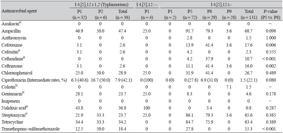
For the antimicrobial resistance analysis presented in Table 3, resistance rates between P5 and P8 isolates were compared using the chi-square test or Fisher’s exact test, as appropriate. Statistical analyses were performed using SPSS version 29.0 (IBM Corp.). Statistical significance was defined as a two-tailed P-value < 0.05.

Table 3. Antimicrobial resistance and gene patterns of Salmonella Typhimurium variants

Antimicrobial agentI 4,[5],12:i:1,2 (Typhimurium) I 4,[5],12:-:- I 4,[5],12:i:-
P1
(n = 32)
P2
(n = 6)
Total
(n = 38)
 P1
(n = 4)
 P1
(n = 2)
P5
(n = 72)
P8
(n = 29)
P9
(n = 28)
Total
(n = 131)
P-value
(P5 vs. P8)
Amikacina)000 0 00000
Ampicillin46.950.047.4 25.0 091.779.33.668.70.098
Azithromycin000 0 02.8001.51.000
Cefotaxime3.102.6 0 013.941.43.617.60.006
Cefoxitina)3.102.6 0 04.2002.30.555
Ceftazidimea)000 0 04.237.9010.7< 0.001
Ceftriaxone3.102.6 0 011.141.43.616.00.002
Chloramphenicol25.050.028.9 25.0 031.941.4026.70.489
Ciprofloxacin (Intermediate rates, %)6.3 (40.6)16.7 (50.0)7.9 (42.1) 0 (100) 0 (0)0 (27.8)6.9 (31.0)0 (0)1.5 (22.1)0.080
Colistinb)000 0 0007.11.5
Gentamicina)28.1023.7 25.0 08.3004.60.178
Imipenem000 0 00000
Nalidixic acida)43.8036.8 100 003.400.80.287
Streptomycina)21.933.323.7 25.0 086.179.33.665.60.385
Tetracycline34.433.334.2 0 084.775.9063.40.389
Trimethoprim–sulfamethoxazole12.550.018.4 0 027.80015.3< 0.001

Values are presented as percentages of resistant isolates unless otherwise indicated.
a)Clinical and Laboratory Standards Institute (CLSI) breakpoints have not been established for these antimicrobial agents; the interpretive standards used are the National Antimicrobial Resistance Monitoring System (NARMS)-established breakpoints for resistance monitoring, which should not be used to predict clinical efficacy. 
b)European Committee on Antimicrobial Susceptibility Testing, 2026.

Results

Distribution of antigenic formulas and gene patterns

Among 173 S. Typhimurium variant isolates, the monophasic variant I 4,[5],12:i:- (n = 131, 75.7%) was the most prevalent, followed by typical S. Typhimurium I 4,[5],12:i:1,2 (n = 38, 22.0%), and the non-motile variant I 4,[5],12:-:- (n = 4, 2.3%). The monophasic variant I 4,[5],12:-:1,2 was not detected.

Gene pattern analysis revealed five patterns (P1, P2, P5, P8, and P9) based on the presence or absence of seven target genes (STM2757, mdh, intergenic regions of fliAfliB, fliC, fljB, fljA, and hin) (Table 2). P5 was the most prevalent (n = 72), followed by P1 (n = 38), P8 (n = 29), P9 (n = 28), and P2 (n = 6).

P1 included 32 typical S. Typhimurium isolates, four I 4,[5],12:-:- isolates, and two I 4,[5],12:i:- isolates. P2 exclusively consisted of six typical S. Typhimurium isolates. P5, P8, and P9 were only detected in I 4,[5],12:i:- isolates. Both P5 and P8 lacked fljA, fljB, and hin, whereas P8 additionally lacked STM2757. P9 was positive for hin, but lacked fljA, fljB, and STM2757.

Among the I 4,[5],12:i:- isolates, 77.1% were classified as Spanish clones (P5, 55.0%; P8, 22.1%), whereas 21.4% corresponded to a U.S. clone (P9).

Antimicrobial resistance profiles

Resistance to ampicillin, streptomycin, and tetracycline was numerically more frequent in P5 and P8 than in the other gene patterns. The resistance rates at P5 and P8 were as follows: ampicillin (91.7% and 79.3%); streptomycin (86.1% and 79.3%); and tetracycline (84.7% and 75.9%), respectively (Table 3).

P8 showed significantly higher resistance to third-generation cephalosporins compared with that of P5 (cefotaxime: 41.4% vs. 13.9%; ceftazidime: 37.9% vs. 4.2%; ceftriaxone: 41.4% vs. 11.1%; P < 0.05). Among 72 P5 isolates, 23 (31.9%) were concurrently resistant to ampicillin, chloramphenicol, streptomycin, and tetracycline, corresponding to four of the five components of the characteristic ACSSuT resistance profile of the Spanish clone. Because sulfamethoxazole alone was not tested, the complete ACSSuT phenotype could not be determined. Twenty-three P5 isolates were resistant to four ACSSuT components (ampicillin, chloramphenicol, streptomycin, and tetracycline) and 18 of these were additionally resistant to SXT.

Typical S. Typhimurium (P1 and P2) and I 4,[5],12:-:- (P1) strains demonstrated high rates of intermediate resistance to ciprofloxacin (42.1% and 100%, respectively). Further, high rates of nalidixic acid resistance were observed in P1 isolates of typical S. Typhimurium (43.8%) and I 4,[5],12:-:- (100%).

In contrast, P9 isolates showed low rates of resistance to most antimicrobials, with resistance to colistin observed in 7.1% of isolates and resistance to other antimicrobials occurring at rates below 4%.

ACT/S-resistant isolates

Overall, 25 isolates (14.5%) were classified as ACT/S-resistant isolates. These included seven typical S. Typhimurium isolates (P1, n = 4; P2, n = 3) and 18 I 4,[5],12:i:- isolates (all P5). No ACT/S-resistant isolates were identified among the P9 strains.

Discussion

Among the isolates collected in South Korea during the study period, I 4,[5],12:i:- was more frequently identified compared with typical S. Typhimurium. P5, P8, and P9 were the predominant gene patterns among I 4,[5],12:i:- isolates, with P5 and P8 previously associated with the Spanish clone and P9 with the U.S. clone. These gene patterns showed substantially different antimicrobial resistance profiles, with P8 isolates showing particularly high resistance to third-generation cephalosporins. These findings reveal the molecular epidemiological diversity of I 4,[5],12:i:- and the association between gene patterns and antimicrobial resistance profiles.

Salmonella serotypes are determined using a combination of somatic (O) and flagellar (H) antigens. Phase variation in S. Typhimurium is regulated by the coordinated expression of two flagellar proteins, FliC (phase 1 antigen) and FljB (phase 2 antigen). Expression switching occurs through inversion of the promoter region, which is mediated by the DNA invertase Hin. This results in fliC suppression by fljA and the subsequent expression of fljB [17, 18]. Gene disruption or deletion within the fljAB operon or associated regulatory regions can result in monophasic variants.

Salmonella Typhimurium (I 4,[5],12:i:1,2) expresses both phase 1 and phase 2 H antigens. However, based on differences in H antigen expression, three variants have been described: monophasic variant I 4,[5],12:i:-, the uncommon variant I 4,[5],12:-:1,2, and the non-motile variant I 4,[5],12:-:- [1921].

The Centers for Disease Control and Prevention and the EFSA report that I 4,[5],12:i:- ranks among the top five most-detected serotypes in human clinical isolates. The I 4,[5],12:i:- variant is increasingly reported in North America, Europe, Asia, Oceania, and South America and is frequently associated with pork, poultry, and cattle products [6, 2228].

A previous nationwide human surveillance in South Korea (2016–2017) reported that I 4,[5],12:i:- accounted for 16.7% of the isolates, surpassing the typical Typhimurium isolates (9.9%) [5]. In the present study, the ratio of I 4,[5],12:i:- to typical Typhimurium increased from 1.7:1 to 3.4:1, indicating that I 4,[5],12:i:- was more frequently identified than typical Typhimurium during the study period. However, this comparison was limited to these two serotypes and does not indicate predominance among all Salmonella serotypes. A recent nationwide surveillance from 2022 to 2024 identified S. Enteritidis (29.7%) as the most common serotype, whereas I 4,[5],12:i:- accounted for 10.5% of the isolates [29].

To improve S. Typhimurium variant identification, the EFSA has proposed molecular approaches targeting the phase 1 and phase 2 flagellar genes. Tennant et al. [8] analyzed genetic variations in fljB, the fliBfliA intergenic region, and the phase 1 “i” antigen gene using PCR-based methods. However, inconsistent findings, including the unexpected absence or presence of certain flagella-associated genes, were occasionally observed. Subsequently, Bugarel et al. [9] proposed a refined PCR-based classification system defining 11 gene patterns (P1–P11) using additional markers, such as fljA, fliC, hin, STM2757, and mdh. This approach enabled improved molecular epidemiologic investigations and facilitated the differentiation of epidemic clones, including the Spanish (P5 and P8) and U.S. (P7 and P9) clones of I 4,[5],12:i:-.

In terms of clonal lineages, I 4,[5],12:i:- has been categorized into several epidemic lineages, including Spanish, U.S., and European clones [19, 27, 28, 30]. In the present study, most isolates showed P5 and P8 patterns, which have been associated with the Spanish clone, whereas P9 has been associated with the U.S. clone. However, as the P1–P11 classification is based on a limited set of PCR markers, these patterns do not allow definitive assignment of isolates to specific clonal lineages. In particular, presence or absence of the European clone, characterized by features such as ST34 and the ASSuT resistance phenotype, could not be determined using the methods employed in this study.

The Spanish monophasic clone, first reported in Spain in 1997, was predominantly associated with phage type U302 and a multidrug resistance profile including resistance to ampicillin, chloramphenicol, sulfonamides, gentamicin, streptomycin, tetracycline, and SXT [9, 31]. This clone is distinct from the classical multidrug-resistant S. Typhimurium DT104 lineage that emerged in the mid-1980s and is characterized by the ACSSuT resistance profile [32]. In the current study, 23 of 72 P5 isolates (31.9%) were resistant to four of the five ACSSuT components (ampicillin, chloramphenicol, streptomycin, and tetracycline) and 18 of these isolates were additionally resistant to SXT. However, because sulfamethoxazole alone was not tested, the complete ACSSuT phenotype could not be determined. These findings should therefore be interpreted as overlapping resistance characteristics rather than evidence of the classical ACSSuT phenotype. Interestingly, P8 isolates, although genetically related to the Spanish clone, showed markedly higher rates of resistance to third-generation cephalosporins than those of P5 isolates. This may limit the treatment options for invasive infections and pose critical clinical and public health challenges. This finding suggests the possible acquisition of additional resistance determinants, underscoring the need for continuous monitoring.

In contrast, the U.S. clone (P9) exhibits low levels of antimicrobial resistance [7]. Consistent with previous reports, the P9 isolates in this study showed low resistance rates, and no ACT/S-resistant isolates were identified. However, detection of colistin resistance in a subset of P9 isolates warrants further investigation into the genetic mechanisms underlying this phenotype.

Overall, three major gene patterns (P5, P8, and P9), previously associated with the Spanish and U.S. clones, were identified among the I 4,[5],12:i:- isolates in South Korea. Predominance of the Spanish clone suggests potential transmission through livestock reservoirs, particularly pork and poultry, as observed in Europe and North America. This finding raises concerns regarding foodborne transmission and highlights the need for integrated surveillance across the food production chain. Furthermore, significant differences in antimicrobial resistance profiles according to gene patterns highlight the epidemiological importance of molecular subtyping.

Considering the increasing prevalence of I 4,[5],12:i:- and its association with the ACT/S resistance phenotype, continued molecular surveillance is essential. Gene pattern-based subtyping may serve as a practical tool for epidemiological investigations and the development of antimicrobial treatment strategies. Overall, these findings highlight the importance of continuous national surveillance under the One Health framework and support the implementation of genomic surveillance and antimicrobial stewardship programs for mitigating the spread of high-risk clones.

Limitations

This study has a few limitations. First, isolates were collected only from university hospitals, which may not fully represent the nationwide epidemiology. Second, whole-genome sequencing was not performed to confirm the clonal relationships and resistance determinants.

Conclusion

Monophasic S. Typhimurium I 4,[5],12:i:- was more frequently identified than typical S. Typhimurium among the isolates included in this study. Most I 4,[5],12:i:- isolates presented gene patterns previously associated with the Spanish clone, whereas a small proportion showed patterns associated with the U.S. clone. Distinct gene patterns were associated with pronounced differences in antimicrobial resistance profiles; particularly, the P8 isolates demonstrated increased resistance to third-generation cephalosporins. These findings highlight the epidemiological and public health relevance of I 4,[5],12:i:- and the importance of continued surveillance. Moreover, they indicate the practicality of gene pattern-based molecular subtyping for epidemiological surveillance and antimicrobial resistance monitoring in resource-limited settings. Overall, this study provides important evidence for public health policies and infection control strategies.

Ethics statement

This study was approved by the Institutional Review Board of Inje University Busan Paik Hospital, with a waiver of informed consent (Approval No. BPIRB NON2024-001).

Conflicts of interest

No potential conflicts of interest relevant to this article were reported.

Funding

The research was supported by a fund (2020E540600) from the Research Program of Korea Disease Control and Prevention Agency and a grant of the Korea Health Technology R&D Project through the Korea Health Industry Development Institute (KHIDI), Ministry of Health & Welfare, Korea (grant number: HR21C1003).

Data availability

Data are available upon reasonable request.

References

1. Popa GL and Papa MI. Salmonella spp. infection – a continuous threat worldwide. Germs 2021;11:88-96.

2. Brenner FW, Villar RG, Angulo FJ, Tauxe R, Swaminathan B. Salmonella nomenclature. J Clin Microbiol 2000;38:2465-7.

3. Jones TF, Ingram LA, Cieslak PR, Vugia DJ, Tobin-D’Angelo M, Hurd S, et al. Salmonellosis outcomes differ substantially by serotype. J Infect Dis 2008;198:109-14.

4. Colarusso G, Peruzy MF, Mazzone P, Ambrosio RL, Pellicanò R, D’Argenzio A, et al. Surveillance of human cases of salmonellosis, campylobacteriosis, listeriosis, and hepatitis A in Campania (Southern Italy): seven-year monitoring (2013-2019). Pathogens 2023;12:48.

5. Kim SH, Sung GH, Park EH, Hwang IY, Kim GR, Song SA, et al. Serotype distribution and antimicrobial resistance of Salmonella isolates in Korea between 2016 and 2017. Ann Lab Med 2022;42:268-73.

6. Qin X, Yang M, Cai H, Liu Y, Gorris L, Aslam MZ, et al. Antibiotic resistance of Salmonella Typhimurium monophasic variant 1,4,[5],12:i:- in China: a systematic review and meta-analysis. Antibiotics (Basel) 2022;11:532.

7. Arai N, Sekizuka T, Tamamura Y, Tanaka K, Barco L, Izumiya H, et al. Phylogenetic characterization of Salmonella enterica serovar Typhimurium and its monophasic variant isolated from food animals in Japan revealed replacement of major epidemic clones in the last 4 decades. J Clin Microbiol 2018;56:e01758-17.

8. Tennant SM, Diallo S, Levy H, Livio S, Sow SO, Tapia M, et al. Identification by PCR of non-typhoidal Salmonella enterica serovars associated with invasive infections among febrile patients in Mali. PLoS Negl Trop Dis 2010;4:e621.

9. Bugarel M, Vignaud ML, Moury F, Fach P, Brisabois A. Molecular identification in monophasic and nonmotile variants of Salmonella enterica serovar Typhimurium. Microbiologyopen 2012;1:481-9.

10. Turner P, Fox-Lewis A, Shrestha P, Dance DAB, Wangrangsimakul T, Cusack TP, et al. Microbiology Investigation Criteria for Reporting Objectively (MICRO): a framework for the reporting and interpretation of clinical microbiology data. BMC Med 2019;17:70.

11. Issenhuth-Jeanjean S, Roggentin P, Mikoleit M, Guibourdenche M, de Pinna E, Nair S, et al. Supplement 2008-2010 (no. 48) to the White-Kauffmann-Le Minor scheme. Res Microbiol 2014;165:526-30.

12. Amavisit P, Boonyawiwat W, Bangtrakulnont A. Characterization of Salmonella enterica serovar Typhimurium and monophasic Salmonella serovar 1,4,[5],12:i:- isolates in Thailand. J Clin Microbiol 2005;43:2736-40.

13. CLSI. Performance standards for antimicrobial susceptibility testing. 36th ed. CLSI supplement M100. Clinical and Laboratory Standards Institute; 2026.

14. Karp BE, Tate H, Plumblee JR, Dessai U, Whichard JM, Thacker EL, et al. National antimicrobial resistance monitoring system: two decades of advancing public health through integrated surveillance of antimicrobial resistance. Foodborne Pathog Dis 2017;14:545-57.

15. EUCAST. Breakpoint tables for interpretation of MICs and zone diameters. Version 16.0. 2026. https://www.eucast.org/clinical_breakpoints [Online] (last visited on 1 June 2026).

16. Crump JA, Sjölund-Karlsson M, Gordon MA, Parry CM. Epidemiology, clinical presentation, laboratory diagnosis, antimicrobial resistance, and antimicrobial management of invasive Salmonella infections. Clin Microbiol Rev 2015;28:901-37.

17. Yamamoto S and Kutsukake K. FljA-mediated posttranscriptional control of phase 1 flagellin expression in flagellar phase variation of Salmonella enterica serovar Typhimurium. J Bacteriol 2006;188:958-67.

18. Switt AI, Soyer Y, Warnick LD, Wiedmann M. Emergence, distribution, and molecular and phenotypic characteristics of Salmonella enterica serotype 4,5,12:i:-. Foodborne Pathog Dis 2009;6:407-15.

19. Sun H, Wan Y, Du P, Bai L. The epidemiology of monophasic Salmonella Typhimurium. Foodborne Pathog Dis 2020;17:87-97.

20. Branchu P, Bawn M, Kingsley RA. Genome variation and molecular epidemiology of Salmonella enterica serovar Typhimurium pathovariants. Infect Immun 2018;86:e00079-18.

21. Arrieta-Gisasola A, Atxaerandio-Landa A, Garrido V, Grillo MJ, Martinez-Ballesteros I, Laorden L, et al. Genotyping study of Salmonella 4,[5],12:i:- monophasic variant of serovar Typhimurium and characterization of the second-phase flagellar deletion by whole genome sequencing. Microorganisms 2020;8:2049.

22. Proroga YTR, Capuano F, Carullo MR, La Tela I, Capparelli R, Barco L, et al. Characterization of Salmonella Typhimurium and its monophasic variant 1,4,[5],12:i:- isolated from different sources. Folia Microbiol (Praha) 2019;64:711-8.

23. Cevallos-Almeida M, Figueroa A, Rojas D, López S, Montalvo T, Llorente P, et al. Colonization of pigs experimentally infected with a monophasic variant of Salmonella Typhimurium. Foodborne Pathog Dis 2018;15:576-82.

24. Merlotti A, Manfreda G, Munck N, Hald T, Litrup E, Nielsen EM, et al. Network approach to source attribution of Salmonella enterica serovar Typhimurium and its monophasic variant. Front Microbiol 2020;11:1205.

25. Canning M, Birhane MG, Dewey-Mattia D, Lawinger H, Cote A, Gieraltowski L. Salmonella outbreaks linked to beef, United States, 2012-2019. J Food Prot 2023;86:100071.

26. Trachsel JM, Bearson BL, Brunelle BW, Bearson SMD. Relationship and distribution of Salmonella enterica serovar I 4,[5],12:i:- strain sequences in the NCBI Pathogen Detection database. BMC Genomics 2022;23:268.

27. Cuenca-Arias P, Montaño LA, Villalobos-Rodríguez AP, Wiesner M, Hidalgo AM. Molecular and phenotypic characterization of Salmonella Typhimurium monophasic variant (1,4,[5],12:i:-) from Colombian clinical isolates. Biomedica 2020;40:722-33.

28. Vazquez X, García V, Fernández J, Rodicio R, Rodicio MR. Insights into the evolution of IncR plasmids found in the Southern European clone of the monophasic variant of Salmonella enterica serovar Typhimurium. Antibiotics (Basel) 2024;13:334.

29. Jeong HJ, Shin E, Kim J, Yoo J. Trends in the serotype distribution of Salmonella enterica isolated from patients with diarrhea in the Republic of Korea from 2022 to 2024. Jugan Geongang Gwa Jilbyeong 2025;18:1790-812.

30. Mourao J, Machado J, Novais C, Antunes P, Peixe L. Characterization of the emerging clinically-relevant multidrug-resistant Salmonella enterica serotype 4,[5],12:i:- (monophasic variant of S. Typhimurium) clones. Eur J Clin Microbiol Infect Dis 2014;33:2249-57.

31. de la Torre E, Zapata D, Tello M, Mejía W, Frías N, García Peña FJ, et al. Several Salmonella enterica subsp. enterica serotype 4,5,12:i:- phage types isolated from swine samples originate from serotype Typhimurium DT U302. J Clin Microbiol 2003;41:2395-400.

32. Carattoli A, Tosini F, Visca P. Multidrug-resistant Salmonella enterica serotype Typhimurium infections. N Engl J Med 1998;339:921-2.
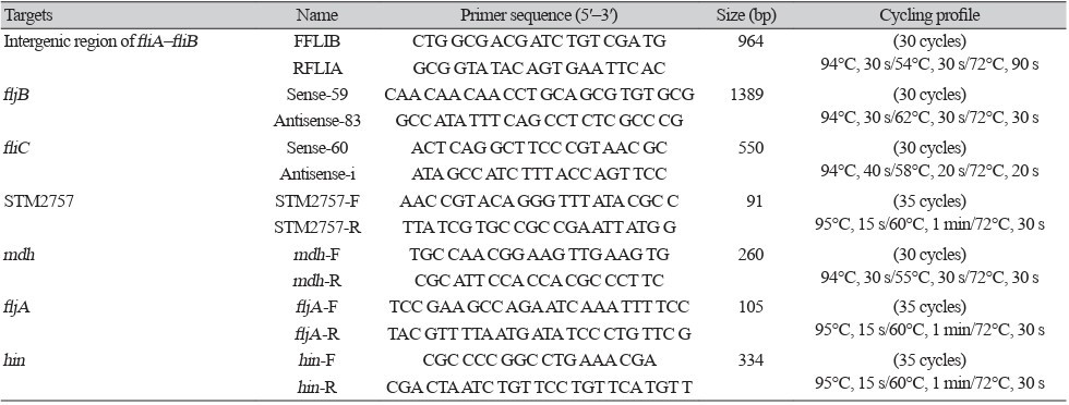
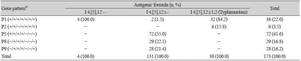
Table 1
Table 2
Table 3

1. Popa GL and Papa MI. Salmonella spp. infection – a continuous threat worldwide. Germs 2021;11:88-96.

2. Brenner FW, Villar RG, Angulo FJ, Tauxe R, Swaminathan B. Salmonella nomenclature. J Clin Microbiol 2000;38:2465-7.

3. Jones TF, Ingram LA, Cieslak PR, Vugia DJ, Tobin-D’Angelo M, Hurd S, et al. Salmonellosis outcomes differ substantially by serotype. J Infect Dis 2008;198:109-14.

4. Colarusso G, Peruzy MF, Mazzone P, Ambrosio RL, Pellicanò R, D’Argenzio A, et al. Surveillance of human cases of salmonellosis, campylobacteriosis, listeriosis, and hepatitis A in Campania (Southern Italy): seven-year monitoring (2013-2019). Pathogens 2023;12:48.

5. Kim SH, Sung GH, Park EH, Hwang IY, Kim GR, Song SA, et al. Serotype distribution and antimicrobial resistance of Salmonella isolates in Korea between 2016 and 2017. Ann Lab Med 2022;42:268-73.

6. Qin X, Yang M, Cai H, Liu Y, Gorris L, Aslam MZ, et al. Antibiotic resistance of Salmonella Typhimurium monophasic variant 1,4,[5],12:i:- in China: a systematic review and meta-analysis. Antibiotics (Basel) 2022;11:532.

7. Arai N, Sekizuka T, Tamamura Y, Tanaka K, Barco L, Izumiya H, et al. Phylogenetic characterization of Salmonella enterica serovar Typhimurium and its monophasic variant isolated from food animals in Japan revealed replacement of major epidemic clones in the last 4 decades. J Clin Microbiol 2018;56:e01758-17.

8. Tennant SM, Diallo S, Levy H, Livio S, Sow SO, Tapia M, et al. Identification by PCR of non-typhoidal Salmonella enterica serovars associated with invasive infections among febrile patients in Mali. PLoS Negl Trop Dis 2010;4:e621.

9. Bugarel M, Vignaud ML, Moury F, Fach P, Brisabois A. Molecular identification in monophasic and nonmotile variants of Salmonella enterica serovar Typhimurium. Microbiologyopen 2012;1:481-9.

10. Turner P, Fox-Lewis A, Shrestha P, Dance DAB, Wangrangsimakul T, Cusack TP, et al. Microbiology Investigation Criteria for Reporting Objectively (MICRO): a framework for the reporting and interpretation of clinical microbiology data. BMC Med 2019;17:70.

11. Issenhuth-Jeanjean S, Roggentin P, Mikoleit M, Guibourdenche M, de Pinna E, Nair S, et al. Supplement 2008-2010 (no. 48) to the White-Kauffmann-Le Minor scheme. Res Microbiol 2014;165:526-30.

12. Amavisit P, Boonyawiwat W, Bangtrakulnont A. Characterization of Salmonella enterica serovar Typhimurium and monophasic Salmonella serovar 1,4,[5],12:i:- isolates in Thailand. J Clin Microbiol 2005;43:2736-40.

13. CLSI. Performance standards for antimicrobial susceptibility testing. 36th ed. CLSI supplement M100. Clinical and Laboratory Standards Institute; 2026.

14. Karp BE, Tate H, Plumblee JR, Dessai U, Whichard JM, Thacker EL, et al. National antimicrobial resistance monitoring system: two decades of advancing public health through integrated surveillance of antimicrobial resistance. Foodborne Pathog Dis 2017;14:545-57.

15. EUCAST. Breakpoint tables for interpretation of MICs and zone diameters. Version 16.0. 2026. https://www.eucast.org/clinical_breakpoints [Online] (last visited on 1 June 2026).

16. Crump JA, Sjölund-Karlsson M, Gordon MA, Parry CM. Epidemiology, clinical presentation, laboratory diagnosis, antimicrobial resistance, and antimicrobial management of invasive Salmonella infections. Clin Microbiol Rev 2015;28:901-37.

17. Yamamoto S and Kutsukake K. FljA-mediated posttranscriptional control of phase 1 flagellin expression in flagellar phase variation of Salmonella enterica serovar Typhimurium. J Bacteriol 2006;188:958-67.

18. Switt AI, Soyer Y, Warnick LD, Wiedmann M. Emergence, distribution, and molecular and phenotypic characteristics of Salmonella enterica serotype 4,5,12:i:-. Foodborne Pathog Dis 2009;6:407-15.

19. Sun H, Wan Y, Du P, Bai L. The epidemiology of monophasic Salmonella Typhimurium. Foodborne Pathog Dis 2020;17:87-97.

20. Branchu P, Bawn M, Kingsley RA. Genome variation and molecular epidemiology of Salmonella enterica serovar Typhimurium pathovariants. Infect Immun 2018;86:e00079-18.

21. Arrieta-Gisasola A, Atxaerandio-Landa A, Garrido V, Grillo MJ, Martinez-Ballesteros I, Laorden L, et al. Genotyping study of Salmonella 4,[5],12:i:- monophasic variant of serovar Typhimurium and characterization of the second-phase flagellar deletion by whole genome sequencing. Microorganisms 2020;8:2049.

22. Proroga YTR, Capuano F, Carullo MR, La Tela I, Capparelli R, Barco L, et al. Characterization of Salmonella Typhimurium and its monophasic variant 1,4,[5],12:i:- isolated from different sources. Folia Microbiol (Praha) 2019;64:711-8.

23. Cevallos-Almeida M, Figueroa A, Rojas D, López S, Montalvo T, Llorente P, et al. Colonization of pigs experimentally infected with a monophasic variant of Salmonella Typhimurium. Foodborne Pathog Dis 2018;15:576-82.

24. Merlotti A, Manfreda G, Munck N, Hald T, Litrup E, Nielsen EM, et al. Network approach to source attribution of Salmonella enterica serovar Typhimurium and its monophasic variant. Front Microbiol 2020;11:1205.

25. Canning M, Birhane MG, Dewey-Mattia D, Lawinger H, Cote A, Gieraltowski L. Salmonella outbreaks linked to beef, United States, 2012-2019. J Food Prot 2023;86:100071.

26. Trachsel JM, Bearson BL, Brunelle BW, Bearson SMD. Relationship and distribution of Salmonella enterica serovar I 4,[5],12:i:- strain sequences in the NCBI Pathogen Detection database. BMC Genomics 2022;23:268.

27. Cuenca-Arias P, Montaño LA, Villalobos-Rodríguez AP, Wiesner M, Hidalgo AM. Molecular and phenotypic characterization of Salmonella Typhimurium monophasic variant (1,4,[5],12:i:-) from Colombian clinical isolates. Biomedica 2020;40:722-33.

28. Vazquez X, García V, Fernández J, Rodicio R, Rodicio MR. Insights into the evolution of IncR plasmids found in the Southern European clone of the monophasic variant of Salmonella enterica serovar Typhimurium. Antibiotics (Basel) 2024;13:334.

29. Jeong HJ, Shin E, Kim J, Yoo J. Trends in the serotype distribution of Salmonella enterica isolated from patients with diarrhea in the Republic of Korea from 2022 to 2024. Jugan Geongang Gwa Jilbyeong 2025;18:1790-812.

30. Mourao J, Machado J, Novais C, Antunes P, Peixe L. Characterization of the emerging clinically-relevant multidrug-resistant Salmonella enterica serotype 4,[5],12:i:- (monophasic variant of S. Typhimurium) clones. Eur J Clin Microbiol Infect Dis 2014;33:2249-57.

31. de la Torre E, Zapata D, Tello M, Mejía W, Frías N, García Peña FJ, et al. Several Salmonella enterica subsp. enterica serotype 4,5,12:i:- phage types isolated from swine samples originate from serotype Typhimurium DT U302. J Clin Microbiol 2003;41:2395-400.

32. Carattoli A, Tosini F, Visca P. Multidrug-resistant Salmonella enterica serotype Typhimurium infections. N Engl J Med 1998;339:921-2.