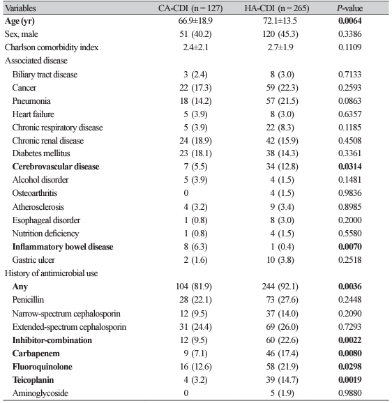
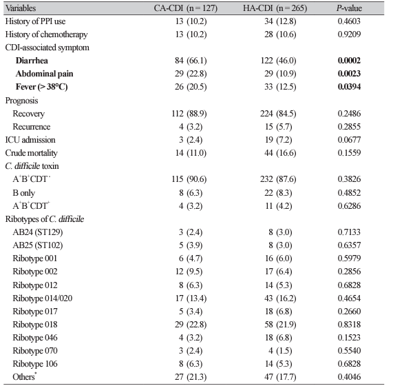
1. Bartlett JG. Clostridium difficile: history of its role as an enteric pathogen and the current state of knowledge about the organism. Clin Infect Dis 1994;18:S265-72. 

2. Wamy M, Pe´pin J, Fang A, Killgore G, Thompson A, Brazier J, et al. Toxin production by an emerging strain of Clostridium difficile associated with outbreaks of severe disease in North America and Europe. Lancet 2005;366:1079–84.
3. Byun JH, Kim H, Kim JL, Kim D, Jeong SH, Shin JH, et al. A nationwide study of molecular epidemiology and antimicrobial susceptibility of Clostridioides difficile in South Korea. Anaerobe 2019;60:102106.
4. Evans ME, Simbartl LA, Kralovic SM, Jain R, Roselle GA. Clostridium difficile infections in Veterans Health Administration acute care facilities. Infect Control Hosp Epidemiol 2014; 5:1037–42.
5. Griffiths D, Fawley W, Kachrimanidou M, Bowden R, Crook DW, Fung R, et al. Multilocus sequence typing of Clostridium difficile. J Clin Microbiol 2010;48:770-8. 
6. McLure A, Clements ACA, Kirk M, Glass K. Modelling diverse sources of Clostridium difficile in the community: importance of animals, infants and asymptomatic carriers. Epidemiol Infect 2019;147:e152, 1–9.
7. McLure A, Clements ACA, Kirk M, Glass K. Clostridium difficile classification overestimates hospital-acquired infections. J Hosp Infect 2018;99:453-60.
8. McDonald LC, Coignard B, Dubberke E, Song X, Horan T, Kutty PK, et al. Recommendations for surveillance of Clostridium difficile-associated disease. Infect Control Hosp Epidemiol 2007;28:140-5.
9. McDonald LC, Gerding DN, Johnson S, Bakken JS, Carroll KC, Coffin SE, et al. Clinical practice guidelines for Clostridium difficile infection in adults and children: 2017 update by the Infectious Diseases Society of America (IDSA) and Society for Healthcare Epidemiology of America (SHEA). Clin Infect Dis 2018;66:e1-48.
10. Guh AY, Adkins SH, Li Q, Bulens SN, Farley MM, Smith Z, et al. Risk factors for communityassociated Clostridium difficile infection in adults: a case-control study. Open Forum Infect Dis 2017;4:ofx171.
11. Kim YA, Park YS, Youk T, Lee H, Lee K. Changes in antimicrobial usage patterns in Korea: 12-year analysis based on database of the National Health Insurance Service-National Sample Cohort. Sci Rep 2018;8:12210.